

Microbiological and Randomly Amplified Polymorphic DNA (RAPD) Marker Protocol for Silicone Condom Lubricant Isolates
- Department of Microbiology, Babcock University
- Department of Microbiology, Faculty of Science, Adekunle Ajasin University, Akungba Akoko. Ondo State Nigeria
- Department of Microbiology, Faculty of Sciences, Adeleke University, Osun Nigeria
Abstract
Condoms serve as a method of birth control. During sexual activity, silicon condom lubricant lowers friction and the chances of harm. Additionally, it increases the protection against STIs, including HIV, by decreasing the likelihood that they may break or fall off. The aim of this study was to perform microbiological and molecular assessments of silicon condom lubricant using a 16S rRNA molecular sequencing protocol. Silicone condom lubricants were randomly selected and analyzed using standard microbiological methods. The molecular identification of the isolated bacteria was performed using the 16S rRNA sequencing protocol. The fungal isolate compendium was characterized using lactophenol cotton blue staining. Antimicrobial susceptibility testing of the isolated organisms was performed using the modified method described by Kirby-Bauer agar disc diffusion. The growth dynamics and killing time were determined using an ultraviolet spectrophotometer with the addition of ciprofloxacin for bacteria and fluconazole for fungi at 24-hour intervals. The bacterial and fungal counts of the selected condom ranged between 1.6 and 9.0 × 10⁻⁴ cfu/ml and between 1.9 and 7.0 × 10⁷ cfu/ml, respectively. The bacteria isolated were both gram-positive and gram-negative, while the fungal species were Aspergillus niger, Byssoctilamiis nivea, Emericella nidulans, Fusarium poae, Eurotium herbariorium, Aspergillus parasiticus and Fusarium oxysporum. All the organisms showed varied resistance and susceptibility. The findings from this study revealed the presence of pathogenic microorganisms in the selected silicone condom lubricant brands sold in Akungba-Akoko. Further studies should be performed to ensure the safety of the silicon condom brands used.
Introduction
Condoms act as a means of contraception and protection against unintended pregnancies and STIs, including HIV. However, silicon lubricant is a potential source of urinary tract infections and other pathogenic microbes1. The first rubber condom was created in 1855. Over the previous ten years, their use has significantly increased. To prevent fluid (such as blood, menstrual fluid, anal mucous, vaginal mucous, or semen) from passing between a sexual partner and their mucous membrane, condoms work by either blocking or erecting a barrier2. Barrier measures, such as condoms, guard against cervical cancer, sexually transmitted diseases, and HIV3. Polyurethane and latex feedstocks offer the greatest protection against HIV/AIDS and sexually transmitted infections4. Currently, the most efficient (100%) method for preventing HIV and STD infections in sexually active individuals is to use condoms correctly and consistently5. In light of this, the promotion of condom use has also received critical attention in the struggle against the fatal HIV/AIDS pandemic6.
However, excessive and incorrect use of condoms could also result in infection by pathogenic organisms, particularly in females, as well as possible allergic reactions in users. Purchasing substandard condoms will have a serious negative impact on all facets of condom advertising and programming. It is not only a waste of scarce financial resources but also undermines the reputation of the low-cost device that has been shown to help stop the spread of HIV/STIs and unwanted pregnancies7. Studies have reported that the spermicidal lubricant functions as a source of UTIs. The investigation of the microbial quality of male and female condoms, which can be a source of growth for microorganisms, was spurred by the lack of reports on microbiological testing, the presence of lubricant, and the greater occurrence of BV in women who are sexually active8. The silicon condom lubricants of male and female condoms were classified using conventional microbiological methods, such as microscopy, growth on specific media/cultural characteristics, biochemical tests, and antibiotic susceptibility tests. The 16S rRNA molecular sequence protocol was used for molecular identification9.
Random amplified polymorphic DNA (RAPD) is a variant of the polymerase chain reaction (PCR) technique based on the amplification of random fragments of DNA (RAPD)10. This technique utilizes short (5–15 mer) oligonucleotide primers of arbitrary sequence at low annealing temperature that hybridize at the loci distributed at random throughout the genome, allowing the amplification of polymorphic DNA fragments11. RAPD was chosen because it can be used to assess genetic relatedness among species.

Selected condom brands used in this study
There is mounting evidence that intravaginal materials, such as condom lubricants, can lead to pathogenic infection12, injure vaginal and rectal tissues, and promote HIV replication in vitro13. Certain condom lubricants may increase the chance of contracting HIV and other STDs as well as bacterial vaginosis14. These lubricants have the potential to disrupt the pH, hydrogen peroxide, and lactobacilli-based vaginal defense mechanisms. For instance, condom lubricants may increase the pH of the vagina and cause chemical harm12, encouraging the growth of organisms associated with BV and resulting in various skin infections15. The bulk of research on condom use that has been published to date has been on condom lubricant because scientists believe that specific lubricants may be linked to BV and other prevalent illnesses in both males and females16. However, there is currently insufficient information available regarding the global prevalence of harmful microorganisms among condom users.
Numerous microorganisms, such as bacteria, viruses, fungi, and protozoa, are present in the human body. Some of them can be categorized as harmful bacteria, but others can be found as typical skin flora in human skin, such as Bacteroides, Staphylococci, Oropharynx Streptococci, anaerobes, Vagina (), and digestive organs (enteric bacilli). The majority of pathogens are spread by feces, which can result in serious illnesses and disease epidemics. Contact of condoms with dirty or unhygienic objects and surfaces could result in contamination with pathogenic microbes17; equally, defects in processing and manufacturing could result in possible contamination, which could result in infections upon use. Hence, this could increase the risk of contracting an unexpected and unknown pathogenic infection from the use of condoms. Previous studies have suggested that organisms such as sp., and are possible pathogenic contaminants4.
The use of silicon condom lubricant as a way to reduce friction and lessen the risk of injury during sexual intercourse prevents falls or breaks, thereby increasing the protection against STIs, including HIV. However, the increase in the incidence of BV and other sexually transmitted diseases has become a major public health concern. Few studies have evaluated the possibility of infection with pathogenic organisms caused by the use of these products (silicone membrane lubricant). There is a high increase in and interest in the use of condoms today because of the role they play in preventing acquired immunodeficiency syndrome and other sexually transmitted diseases. It is also a commonly used method of contraception. However, there is no information on the microbiological assessment of silicone membranes despite the increase in their usage. Hence, it is of utmost importance that microbiological quality and assessment of silicone condom lubricant be evaluated to establish the safety of silicon condom lubricant.
Materials AND METHODS
Sample collection and preparation
The study adopted an experimental design. The silicone membrane lubricant samples used in this study were randomly selected and purchased from local patents selling medicine and from pharmacy shops in the Akungba-Akoko community, Ondo State, Nigeria. Briefly, five test tubes were filled with nine millilitres of distilled water each, the mouth was sealed with cotton wool and covered with aluminum foil, and the mouth was sterilized by autoclaving for fifteen minutes at 121 degrees Celsius. After sterilization, the water was allowed to cool, and the test tubes containing sterile distilled water were then labeled 10-10. Simultaneously, the condom surfaces were swabped with 75% ethanol, removed aseptically, inserted into 9 ml of sterile distilled water labeled 10, swirled gently and then diluted serially1.
Bacteriological analysis of silicone membrane lubricant samples
Fivefold serial dilutions (0.5 ml) of 10 and 10 condom samples were aliquoted into sterile petri dishes using the pour plate method. The agar used (macConkey agar, plate count agar and blood agar) was prepared based on the manufacturer’s instructions. The plates were sterilized using an autoclave at 121°C for 15 minutes. After sterilization, 0.5 ml of the inoculum was added to the plates, and the cooled media were poured into the plates, mixed evenly and allowed to set. However, the plates were incubated at 37°C for 24 hours, the colonies were counted and recorded accordingly, and the cultural and morphological characteristics of the isolates were observed and recorded. The colonies were subcultured on fresh agar plates, and pure cultures were obtained and preserved on nutrient agar slants and kept in a refrigerator for further studies18, 19.
Microscopy, macroscopic examination and biochemical identification of the isolates
Culture characteristics and microscopic examination were carried out to identify the pure isolates. Preliminary identification of the organisms was based on cellular morphological characterization, including color, size, and colony characteristics (form, margin, and elevation). Grain staining and biochemical tests, such as catalase, oxidase, coagulase, Gram staining, citrate, sugar fermentation (dextrose, sucrose, lactose, starch hydrolysis), motility, indole test, urease, hydrogen sulfide, and gas production, were carried out for conventional identification of the isolates and compared with Bergey’s manual of determinative microbiology for identification18, 20.
Fungal Identification
Colonies of the fungi were subcultured on potato dextrose agar (PDA), and colonies on the plates were observed. The isolates were then further subjected to microscopic observation by staining with two drops of lacto-phenol cotton blue on a glass slide and observed under a microscope using a 40× objective lens21.
Antibacterial susceptibility testing of bacteria isolated from silicone membrane lubricant
Antimicrobial susceptibility testing (AST) was performed using Kirby-Bauer’s disc diffusion method on Muller-Hinton agar22. The purpose of this test was to identify the phenotypic resistance characteristics of the isolated bacteria to commonly prescribed antibiotics. Twenty-four-hour-old slant cultures were inoculated into nutrient agar broth and adjusted to the 0.5 McFarland standard. The standardized inoculum was inoculated on Mueller-Hinton agar plates. The plates were allowed to incubate for 15 minutes to allow the inoculum to diffuse before the antibiotic-impregnated discs (Oxoid) were applied using a pair of sterile forceps. The discs were gently placed and pressed firmly on agar using sterile forceps. The plates were inverted and incubated overnight at 37°C. After 24 hours of incubation, the plates were examined. A clear zone of inhibition was observed around each disc and was recorded and interpreted according to the Clinical Laboratory Standard Institute guidelines23, 24.
Antibacterial susceptibility test of silicone condom lubricant fungal isolates
Five antifungal drugs were used in this study: ketoconazole, nystatin, tinidazole, fluconazole and griseofulvin. The antifungal tablets were ground into a fine powder using a mortar and pestle. The ground tablets were measured at different concentrations: 0.02 g/ml, 0.04 g/ml and 0.06 g/ml25.
Determination of Antibiotic Sensitivity Pattern
The antibiotic sensitivity patterns of the antifungal drugs against the isolated fungi were determined using the agar well diffusion method. This study employed a newly developed agar-based method using the agar well method to determine the susceptibility of yeast isolates to antifungal agents. A standardized concentration of inoculum (10) with a fixed volume (1 ml) was inoculated on Mueller Hinton agar. Different concentrations of antifungal drugs (approximately 1 drop) were then added to the agar wells. The inoculated plates were incubated at 25°C for 72 hours, and the zones of inhibition were determined in millimeters and recorded accordingly26.
Molecular identification using the 16S rRNA sequence protocol
DNA extraction and PCR
The DNA of the isolates was extracted using the Quick-DNA™ Fungal/Bacterial Miniprep Kit (Zymo Research) according to the manufacturer’s instructions. The extracted DNA was then stored at -20°C until PCR analysis26.
PCR sequencing preparation cocktails were prepared using (per reaction) 25 µl of Taq 2X Master Mix (NEB) and 4 µl of 10 pmol each of the forward and reverse primers (27F 5’- AGA GTT TGA TCM TGG CTC AG-3’ and -1525R, 5′-AAGGAGGTGATCCAGCC-3′). Then, 42 µl of sterile distilled water was added to 8 μl of the DNA template. PCR was carried out in a GeneAmp 9700 PCR System Thermocycler (Applied Biosystem Inc., USA) with a profile containing initial denaturation (94°C for 5 min), followed by 30 cycles of 94°C for 30 s, 50°C for 60 s and 72°C for 1 minute 30 seconds and a final termination at 72°C for 10 minutes. The mixture was allowed to chill at 4°C27, 28.
To verify amplification, the integrity of the amplified gene fragment was examined on a 1% agarose gel. After the addition of the buffer (1× TAE buffer), a 1.5% agarose gel was produced. For five minutes, the suspension was boiled in a microwave. After the melted agarose cooled to 60°C, it was stained with 3 µl of 0.5 g/ml ethidium bromide, which converts invisible UV light into visible orange light. Melted agarose was poured into the casting tray by inserting a comb into the tray. To create the wells, the gel was left to solidify for 20 minutes. The gel was barely submerged when 1X TAE buffer was added to the gel tank. Following the loading of the 100 bp DNA ladder into the wells, 4 µl of each PCR product was added to 2 µl (2 µl) of 10X blue gel loading dye, which provides color and density to the samples to facilitate loading into the wells and monitoring the gel's progress. The gel was photographed after 45 minutes of electrophoresis at 120 volts, as observed by UV transillumination. The mobility of a 100 bp molecular weight ladder, which was run alongside experimental samples in the gel, was used to assess the sizes of the PCR products27.
Purification of the amplified product
Once the gel integrity was restored, the amplified fragments were ethanol filtered to eliminate the PCR reagents. Briefly, 240 µl of 95% ethanol and 7.6 µl of 3 M Na acetate were added to each 40 µl of PCR-amplified product, which was transferred to a fresh, sterile 1.5 µl Eppendorf tube. The solution was mixed well by vortexing, and the tube was maintained at -20°C for 30 minutes. The pellets were mixed and washed with 150 µl of 70% ethanol before centrifugation for 15 minutes at 7,500 ×g and 4°C, followed by removal of the supernatant and centrifugation for 10 minutes at 13,000 ×g and 4°C. Once more, the mixture was allowed to dry in the fume hood for ten to fifteen minutes at ambient temperature after the supernatant was removed. The sample was then resuspended in 20 µl of sterile distilled water and stored at -20°C before sequencing. A 1.5% agarose gel run at 110 V for approximately one hour was used to confirm the presence of the purified product, and a NanoDrop Thermo Scientific Model 2000 was used for quantification28.
Molecular sequencing of bacterial isolates
Using the Big Dye Terminator v3.1 cycle sequencing kit, the amplified fragments were sequenced using an Applied Biosystems Genetic Analyzer 3130xl sequencer following the manufacturer's instructions. For all genetic studies, MEGA 6 and BioEdit tools were utilized27.
Growth dynamics and death rates of the isolates determined using an ultraviolet spectrophotometer
The term growth dynamic describes how quickly a microorganism's cells increase at a particular point in time. The purpose of this test was to ascertain the growth rate of the isolates and the timing of their death. Colonies were picked from the stocked culture aseptically, placed in nutrient broth and incubated at 37°C for 24 hours. A loopful of the isolates was picked from the culture into nutrient broth in four sets, which were set A, B, C and D. Then, 0.5 ml of ciprofloxacin, sedofloxacin and erythromycin were added to sets A, B, and C, respectively, to determine the killing time, while set D was left to further check the growth rate of the microorganisms using an ultraviolet spectrophotometer set at a wavelength of 620λ, which was warmed for 15 minutes. The control was first read (which was sterile broth with no organisms), and the first reading was taken at zero hours and read at intervals of every 12 hours 8 times29.
Data analysis
The data were analyzed via descriptive analysis, and statistically, using SPSS version 20, the means of the zones of inhibition were determined via one-way ANOVA, and significant differences were considered at p≤ 0.05.
RESULTS AND DISCUSSION
In this study, a total of 13 isolates were recovered from different samples used. The isolated organisms were identified through microscopic examination, preliminary biochemical tests, and sugar fermentation. Molecular identification was further used to identify the selected bacterial and fungal isolates.
Gram reaction and microscopic examination of organisms isolated from silicon condom lubricant
|
Parameter |
Isolate code |
Gram reaction |
Shape |
|
Male Silicone Condom (Expired) |
M-SCL |
+ |
Rod |
|
M-SCL 2 |
+ |
Rod | |
|
GC-SCL |
+ |
Cocci | |
|
GC-SCL 2 |
+ |
Rod | |
|
NV-SCL |
+ |
Cocci | |
|
Male Silicone Condom (New) |
K-SCL |
+ |
Cocci |
|
GC-SCL |
+ |
Rod | |
|
F-SCL |
+ |
Rod | |
|
GC-SCL 2 |
+ |
Cocci | |
|
GC-SCL 3 |
+ |
Cocci | |
|
F-SCL |
- |
Cocci | |
|
GC-SCL |
- |
Vibro | |
|
Female Silicone Condom (New) |
FC-SCL |
+ |
Rod |
Cultural characteristics of fungal isolates obtained from silicon condom lubricant
|
Isolate code |
Growth rate |
Diameter of colony |
Pigmentation |
Hyphae |
Conidum |
|
K-SCL |
Fastidious |
85 mm |
Black conidia |
Cylindrical phialides |
Branched out from the phialide |
|
GC-SCL |
Moderate |
18 mm |
Gray‒Green conidiophores |
Short cylindrical phialides |
Branched out from the phialide |
|
F-SCL |
Fastidious |
46 mm |
Dark-Red‒Brown |
Short conidia head |
Branched out from the phialide |
|
GC-SCL 2 |
Fastidious |
60 mm |
Pale-Brown |
Phialides are usually situated directly on the hyphae |
Branched out from the hyphae |
|
GC-SCL 3 |
Fastidious |
55 mm |
Whitish |
Short phialides |
Phialide are borne from short brown conidiophores |
|
GC-SCL 4 |
Fastidious |
55 mm |
White to peach |
Short and broad mono-phialides |
Branched from the monophialides, not sporodochia |
|
GC-SCL 5 |
Fastidious |
50 mm |
Dark-Yellow Green |
Cylindrical phialides |
Branched out from the phialide |
Bacterial and fungal count silicon condom lubricant cultured on plate count agar, blood agar, and MacConkey agar
|
Agar |
Isolate Code |
Bacterial count (TBC) cfu/ml |
Fungal count (TFC) on PDA agar cfu/ml | ||||
|
Male (new) |
Male (expired) |
Female (New) |
Male (new) |
Male (expired) |
Female (New) | ||
|
Blood Agar |
M-SCL |
- |
5.5 x 10⁻⁴ |
- |
- |
- |
- |
|
GC-SCL |
- |
3.1 x 10⁻⁴ |
- |
2.0 x 10⁷ |
- |
- | |
|
GC-SCL 2 |
6.4 x 10⁻⁴ |
9.0 x 10⁻⁴ |
- |
- |
3.7 x 10⁷ |
- | |
|
K-SCL |
1.6 x 10⁻⁴ |
- |
- |
7.0 x 10⁷ |
- |
- | |
|
GC-SCL 3 |
8.2 x 10⁻⁴ |
- |
- |
- |
1.9 x 10⁷ |
- | |
|
F-SCL 2 |
3.5 x 10⁻⁴ |
- |
- |
- |
- |
- | |
|
GC-SCL 4 |
- |
- |
- |
- |
2.8 x 10⁷ |
- | |
|
FC-SCL |
- |
- |
3.4 x 10⁻⁴ |
2.8 x 10⁷ |
- |
- | |
|
Plate Count Agar |
NV-SCL |
5.6 x 10⁻⁴ |
- |
- | |||
|
GC-SCL |
1.6 x 10⁻⁴ |
- |
- | ||||
|
F-SCL |
3.2 x 10⁻⁴ |
- |
- | ||||
|
GC-SCL 3 |
4.8 x 10⁻⁴ |
- |
- | ||||
|
MacConkey Agar |
M-SCL 2 |
- |
6.0 x 10⁻⁴ |
- | |||
Probability identities of bacteria and fungi from silicon condom lubricant isolates
|
Categories |
Isolates code |
Probable bacteria identified |
Probable fungi identified |
|
Male (Expired) condoms |
M-SCL |
Bacillus species |
- |
|
M-SCL 2 |
Escherichia coli |
- | |
|
GC-SCL |
Staphylococcus species |
Eurotium herbariorum | |
|
GC-SCL 2 |
Escherichia coli |
Byssoctilamis nivea | |
|
GC-SCL 3 |
- |
Fusarium oxysporum | |
|
GC-SCL 4 |
- |
Aspergillus parasiticus | |
|
GC-SCL 5 |
- |
Fusarium poae | |
|
Male (New) condoms |
NV-SCL |
Streptococcus species |
- |
|
K-SCL |
Staphylococcus species |
Aspergillus niger | |
|
GC-SCL |
Bacillus subtilis |
- | |
|
F-SCL |
Bacillus subtilis |
Emericella nidulans | |
|
GC-SCL 2 |
Salmonella species |
- | |
|
GC-SCL 3 |
Staphylococcus species |
- | |
|
F-SCL 2 |
Vibrio species |
- | |
|
GC-SCL 4 |
Staphylococcus sp. |
- | |
|
Female (New) condoms |
FC-SCL |
Aeromonas sp. |
|
Bacterial identity and accession numbers determined using 16S rRNA
|
Sample ID |
Scientific Name |
Max Score |
Total Score |
Query Cover |
E- value |
Perentage Identity |
Accession No. |
|
KISS B.A (5EMKIS) |
|
2756 |
2756 |
100% |
0 |
99.87% |
NR118997 |
|
EXP GOLD B.A (12COF EXPBA) |
|
2724 |
2724 |
100% |
0 |
99.87% |
L37597 |

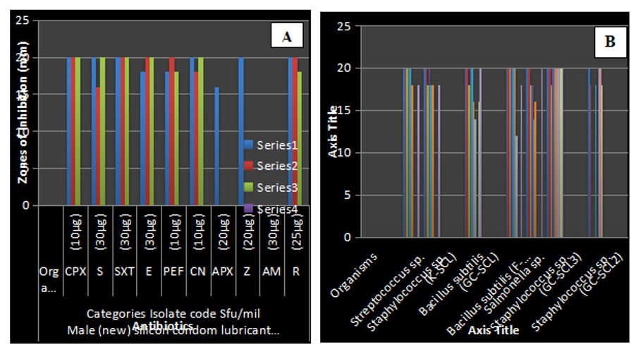
Antibiotic Susceptibility of Gram-Positive Bacteria Isolated from (A) Male-Expired (B) Male (New) Silicon Condom Lubricant (mm) (p≤ 0.05)

Antibiotic-susceptible

Antibiotic susceptibility test of identified gram-negative bacterial isolates from silicon condom lubricant in terms of the diameter of the zones of inhibition. KEY: S- Streptomycin, PN- Ampicillin, CEP- Ceporex, OFX- Tarivid, NA- Nalidixic, PEF- Reflacine, CN- Gentamycin, AU- Augmentin, CPX- Ciproflox, SXT- Septrin

Antifungal susceptibility of fungal isolates obtained from (a) male New (b) Male Expired silicon condom lubricant (mm)

Growth dynamics of bacterial isolates from (a) male-expired, (b) female-new, and (c) male-new silicon condom lubricants (wavelength 620λ)

Killing time of bacteria isolated from a) expired male b) new male c) new female silicon condom lubricant with the addition of ciprofloxacin at 24 h (wavelength 620λ)

Growth dynamics of fungi isolated from male and male expired silicon condom lubricant

Killing time of fungi isolated from male new (A) and male expired (B) silicon condom lubricant samples

Agarose gel showing amplified positive bands (1500 bp) of the selected bacterial isolates and the phylogenetic relatedness of the bacterial isolates (b) and fungal isolates (c). Mk- molecular ladder, Lane 1, 2- bacterial strain showing band at 1500 bp
Figure 4 shows the antibiotic susceptibility of the identified gram-negative organisms. In this table, it was shown that both the isolates had a 20 mm zone of inhibition to Pefloxacin and ciprofloxacin. sp. had the greatest zone of inhibition to streptomycin (20 mm), while had the lowest zone of inhibition to streptomycin (18 mm). Both isolates were resistant to ampicillin, nalidixic acid, Ceporex, and septrin. . and sp. had the greatest zone of inhibition to Augmentin (18 mm). . had the highest zone of inhibition for Tarivid, while the Tarivid Flex BA had the lowest zone of inhibition, as shown in Figure 4.
The antifungal susceptibility test of the identified fungi from the silicon condom lubricant is shown in Figure 5. In this table, it was observed that the abundance of the organism was greatest (36 mm), while had the lowest zone of inhibition (12 mm) to fluconazole. Griseofulvin had the greatest zone of inhibition (32 mm) for B. nivea. had the greatest zone of inhibition (34 mm), while had the lowest zone of inhibition of ketoconazole (15 mm). To our knowledge, had the greatest zone of inhibition (34 mm), while had the lowest zone of inhibition (14 mm).
The growth dynamics of bacteria isolated from the silicon condom lubricant are shown in Figure 6. An ultraviolet spectrophotometer was used at a wavelength of 620λ. At 0 hours, sp. had the highest growth rate of 0.342λ, while sp. had the lowest death rate of 0.123λ. At 64 h, . had the lowest death rate (0.021λ), while had the highest death rate (0.310λ). Figure 7 shows the killing time of the bacterial isolates and the addition of the antibiotic ciprofloxacin at the 24 hour using an ultraviolet spectrophotometer. At 0 h, sp. was killed at 0.342λ, while sp. (0.096λ) was killed at 64 h. had the lowest death rate of 0.097λ, while sp. had the highest death rate of 0.289λ.
Figure 8 shows the growth dynamics of the fungal isolates in the samples using an ultraviolet spectrophotometer at a wavelength of 620λ. At 0 h, B. nivea had the highest growth rate (0.322λ), while had the lowest death rate (0.140λ). At 64 h, had the highest growth rate (0.297λ), while had the lowest death rate (0.195λ). Figure 9 shows the killing time of fungal isolates and the addition of fluconazole at 24 hours. However, at 0 h, had the highest mortality rate at 0.250λ, while had the lowest mortality rate at 0.094λ at 64 h; had the highest mortality rate at 0.221λ, while had the lowest mortality rate at 0.095λ.
The bacterial identity, accession number and percentage similarity are shown in
Condoms are the most effective barrier methods for protecting against HIV, STRs and cervical cancer30, 31. The use of low condoms has drawbacks, such as a greater risk of HIV/STD transmission and unintended pregnancies, which increase the possibility of maternal and newborn death32. However, the use of poor-quality condoms has been known to adversely affect condom reliability and safety. Additionally, reports of adverse effects arising from the use of condoms have negatively affected the credibility and acceptability of condoms21. The observed variations in bacterial count among the various brands might influence the variation in the manufacturing dates/shelf life33 of silicon condom lubricants. The presence of gram-negative organisms was an indication that the development of pathogenic infections arising from the use of these products is highly feasible. The culture characteristics of the fungal isolates shown in
The bacterial loads of six popular condom brands are high. The relatively high microbial counts can be due to contaminants during processing, manufacturing, and packaging as well as during isolation33. 22, who reported similar high bacteria counts in the tested silicon condom lubricants, reported that the presence of bacteria in condoms has been a major source of vaginal contamination during sexual intercourse and other STDs, such as Gonorrhoae and Syphillis. A total of thirteen selected bacterial isolates were identified: sp sp sp sp sp sp. The fungal species were and These microorganisms were probably found in the silicon condom lubricants because they derive nutrients and other growth requirements from them. This finding conforms with Bergey’s manual of determinative microbiology, as well as the findings of22, who reported the isolation of similar organisms from condoms. Since it has been reported that under different conditions, bacterial populations exhibit different morphological and physiological characteristics33, determining the pattern of antibiotic susceptibility of isolates is therefore crucial. The susceptibility of the identified gram-positive bacteria to the selected antibacterial agents showed that sp. isolated from gold circle silicon condom lubricant was the least resistant organism and was susceptible to all antibiotics tested.
However, a second strain of sp. isolated from gold circle silicon membrane lubricant was observed to be the most resistant gram-positive bacteria obtained, showing complete resistance to Ampicillin, Zinnacef, and Amoxicillin while displaying partial resistance to Reflacine and gentamycin. On the other hand, antimicrobial susceptibility patterns of gram-negative isolates revealed that sp. isolated from flex silicon condom lubricant was least susceptible to the tested antibiotics, with complete resistance to Septrin, Ceporex, Ampicillin and Nalidixic. The observed high antimicrobial resistance displayed by some bacterial species can be attributed to the fact that these organisms were possible contaminants that originated from people who had been exposed to a wide variety of antibiotics, either at the production and packaging stages of manufacturing or during the isolation process34. Amoxicillin and Septrin were found to be the least effective antimicrobial agents for gram-positive and gram-negative organisms, respectively. Similarly, tinidazole was the least effective antifungal agent tested, while ketoconazole was the most effective. Generally, isolated organisms were found to be resistant to Ampicillin, Zinnacef, and Amoxicillin, which could result in the development of antimicrobial resistance when used to treat infected individuals.
An ultraviolet spectrophotometer was used to determine the lag phase, log phase, stationary phase, and death phase of the organisms, as well as to determine the death rate of the organisms after the addition of ciprofloxacin. The highest growth of 0.728λ was observed for sp. obtained from the gold (expired) membrane, while the lowest growth of 0.380 λ was observed for sp. isolated from the PCA of New Vietnam at 24 hours. Similarly, after 64 hours, sp. had the highest death rate at 0.110λ, while sp. had the lowest death rate at 0.296. The addition of ciprofloxacin to bacterial isolates was observed to affect their growth dynamics and killing time. After 24 hours, the highest growth rate of 0.628λ was observed for Staphylococcus sp. obtained from the gold (expired) membrane, while isolated from the gold membrane showed the lowest growth rate of 0.405λ. At the 64th hour, sp. had the lowest death rate (0.289λ) when isolated from the gold membrane, and sp. had the highest death rate (0.089λ). The amount of new bacteria that appeared per unit of time in the fungal isolate after the addition of fluconazole for 24 hours was proportional to the starting population. Therefore, if growth is unrestricted, doubling will continue at a steady pace, doubling both the population's growth rate and its number of cells with each passing time interval. Ciprofloxacin was added during the exponential phase to hasten the rate at which the organisms died. This clarifies that the rate at which organisms die can be managed with the use of antibiotics35.
With the development of PCR technology and ongoing improvements in nucleic acid research technology, the 16S rRNA molecular sequence procedure has become the most popular method for identifying and detecting diseases. It also aids in gene discovery35. The 16S rRNA molecular sequence protocol aids the conventional method to establish the accuracy of the findings. The bacteria used were (NR036904 and L37597) and . The findings from this study, therefore, established the presence of pathogenic organisms in condom silicon lubricant brands sold in Akungba, Akoko. The sequencing results of the PCR-amplified 16S rRNA gene revealed that the selected bacterial isolate belonged to the genus . This strain exhibited 99.87% similarity with strain 1 and strain 2 (NR036904 and L37597). The phylogenetic relatedness indicated that the fungus is closely related to . However, strain 1 is closely related to strain 2, and they are closely related to
Conclusion
The presence of pathogenic organisms in the selected silicone condom lubricant brands sold in Akungba-Akoko was high. Although no definite link was established between the occurrence of STDs and the use of silicon membrane lubricant, the presence of certain pathogenic microbes obtained in this study from the selected silicone membrane lubricant could be a major health risk to the users. In this study, proper handling, manufacturing, packaging and proper processing were observed. Proper information and awareness should be provided for people who buy certain silicon condoms lubricant without knowing the validity of the product they use. The indiscriminate use of antibiotics, which leads to antimicrobial resistance of the microorganism obtained from the silicone condom lubricant, should be discouraged. Further studies should be performed on silicone condom lubricant to ascertain the safety of the brands used.
ACKNOWLEDGEMENTS
The authors of this article would like to thank all of the technical personnel for their assistance in the laboratory section of the Department of Microbiology, Faculty of Science, Adekunle Ajasin University, Akungba-Akoko, Ondo State, Nigeria.
Data availability statement
The datasets generated during and/or analyzed during the current study are available from the corresponding author upon reasonable request.
Funding Statement
The authors declare that no funds, grants, or other support was received during the preparation of this manuscript.
Conflict of interest disclosure
The authors declare that there are no conflicts of interest.