

Chemical constituents and bioactivity of Gynura procumbens (Lour.) Merr.
- Faculty of Chemistry, University of Science, VNU-HCM
Abstract
Introduction: Gynura procumbens (Lour.) Merr. (Family: Asteraceae) is mainly popular in South-East Asian countries for its traditional medicinal properties. It is usually used as a traditional medicine for the treatment of eruptive fevers, rash, kidney disease, migraines, constipation, hypertension, diabetes mellitus, and cancer. It is commonly used as a traditional medicine in Vietnam for the treatment of many diseases.
Methods: The leaves and trunks of G. procumbens were collected, macerated with methanol. The extracts from MeOH-soluble extract were processed by the column chromatographic technique to give pure compounds and the nuclear magnetic resonance methods were applied to determine their chemical structures. The inhibitory activities of these extracts against α-glucosidase were conducted and compared with acarbose.
Results: Seven organic compounds were isolated and determined the structures, including syringic acid (1), quercetin (2), N,N-dimethylanthranilic acid (3), dehydrovomifoliol (4), β-sitosterol 3-O-β-D-glucopyranoside (5), schottenol (6), montanic acid (7). The inhibition of α-glucosidase test results the IC50 values of the four extracts which were lower than those of acarbose.
Conclusion: Seven pure compounds were identified from the leaves and trunks of G. procumbens, including two compounds being isolated from G. procumbens for the first time. The test results showed that the the parts of G. procumbens were active as α-glucosidase inhibitor, which would be useful to support the treatment for diabetes.
Introduction
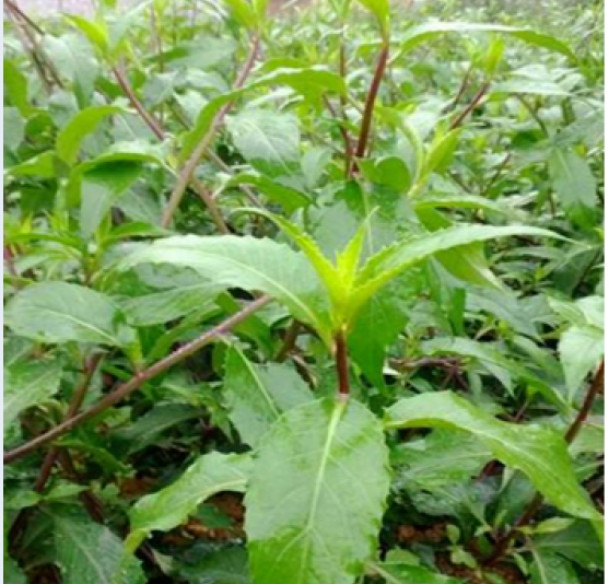
Gynura is the genus of the Asteraceae family, includes 20 species spread all over the world, particularly in Vietnam, China, Malaysia, Thailand, Indonesia, Korea, and the Philippines. G. procumbens (Lour.) Merr. (Figure 1) is a herbal material widely used in tropical countries for the treatment of various health ailments such as cancers, lymphatic pain, hypertension, skin diseases, diabetes mellitus…)2, 1. Nowadays, people in various tropical regions consume an increasing amount of G. procumbens leaves in diet and tea. Research shows that the leaves do not have any toxicity2. Pharmacologic studies have reported that G. procumbens has antioxidant, anti-Herpes simplex, anti-hyperglycemic, anti-hyperlipidemic, anti-inflammatory, analgesic, and reducing blood hypertension properties. The health benefits of G. procumbens are related to some of its bioactive compounds, such as flavonoids, saponins, and alkaloids3. However, there were not many studies about the chemicals constituent of this plant, especially in Vietnam. This study aimed to investigate the chemical constituents from the leaves and trunks of G. procumbens, growing in Gia Lai Province, Vietnam. By column chromatographic and spectroscopic methods seven compounds (1-7) (Figure 2) were identified. Besides, we also tested the inhibitory activity of α-glucosidase on four extracts of this plant.


Chemical structures for1-7.
Methods
Chemicals and equipment
Column chromatography was performed on silica gel (HiMedia) (230-400 Mesh). Thin-layer chromatography (TLC) and preparative TLC were performed on silica gel GF(Merck), visualized by hot 10 % solution of HSO. NMR spectra were acquired on Bruker 500 Avance III at 500 MHz for H-NMR and 125 MHz for C-NMR spectra.
The pure solvents methanol, ethyl acetate, n-butanol, petroleum ether, chloroform were from Chemsol Vina, Vietnam.
Acarbose, an α-glucosidase inhibitor, was from Chem Cruz, Santa Cruz Biotechnology, Inc., USA.
Plant material
The leaves and trunks of G. procumbens were collected at Gia Lai province, Vietnam, in July 2016 and authenticated by Dr. Dang Van Son, Department of Biological Resources, Institute of Tropical Biology – Ho Chi Minh City, Vietnam.
Extraction and isolation
Dried leaves and trunks were ground into powder (2.9 kg) and extracted with hot methanol (4 × 7 L) for four hours each time. The methanolic filtrate was then evaporated to dryness under reduced pressure to obtain a methanolic residue (375.0 g). The methanolic residue was then dissolved in aqueous methanol (10 % methanol) and extracted with petroleum ether (10 x 500 mL), ethyl acetate (10 x 500 mL), n-butanol (10 x 500 mL), consecutively, to afford petroleum ether (PE, 75.0 g), ethyl acetate extract (EA, 7.2 g), n-butanol (Bu, 9.9 g) and crystal compound (102.9 g). The ethyl acetate extract (EA, 7.2 g) was subjected to a silica gel column chromatography and eluted with petroleum ether–ethyl acetate (stepwise, 9:1 → 0:10) followed by ethyl acetate–methanol (stepwise, 8:2 → 6:4) to afford five main fractions EA1 (26.1 mg), EA2 (498.1 mg), EA3 (696.2 mg), EA4 (1200.6 mg), EA5 (627.9 mg). Fraction EA1 (26.1 mg) was washed and cleaned with methanol (MeOH) to give compound 1 (9.0 mg). Fraction EA5 (1200.6 mg) was subjected to a silica gel column chromatography, eluted with chloroform–methanol (CHCl–MeOH) (stepwise, 99:1 → 9:1) to give compound 3 (11.0 mg). Fraction EA3 (627.9 mg) was subjected to column chromatographic separation over silica gel and eluted with CHCl–MeOH (stepwise, 99:1 → 9:1) to give compound 2 (4.4 mg). The same manner was applied on the EA2 (498.1 mg), eluted with CHCl–MeOH (95:5) to give compound 4 (4.5 mg). Fraction EA4 (696.2 mg) was fractionated by a silica gel column chromatography using CHCl–MeOH (stepwise, 95:5 → 8:2) to give compound 5 (4.5 mg). The petroleum ether extract (PE, 75.0 g) was subjected to a silica gel column chromatography and eluted with petroleum ether–ethyl acetate (PE–EA) (stepwise, 9:1 → 0:10) to afford fractions, in these, there were two fractions which were coded as PE1 (1294.7 mg), PE2 (2851.0 mg). By subjecting to a silica gel column chromatographic and eluting with appropriate solvents, fraction PE1 gave compound 6, PE2 gave compound 7.
Compound 1 (syringic acid): white needle-shaped crystals, H- and C-NMR (
Compound 2 (quercetin): yellow powder, H- and C-NMR (
Compound 3 (N,N-dimethylanthranilic acid): white powder, H- and C-NMR (
Compound 4 (dehydrovomifoliol): white crystals, H- and C-NMR (
Compound 5 (β-sitosterol 3-O-β-D-glucopyranoside): white powder, H-NMR (pyridine-d) δ(ppm): 3.93 (1H, m, H-3),5.34 (1H, m, H-6), 0.65 (3H, s, H-18), 0.92 (3H, s, H-19), 0.98 (3H, d, J = 6.5, H-21), 0.85 (3H, d, J = 7.0, H-26), 0.87 (3H, d, J = 7.0, H-27), 0.88 (3H, t, J = 7.0, H-29) C-NMR (pyridine-d) δ (ppm): 37.7 (C-1), 30.4 (C-2), 78.7 (C-3), 40.1 (C-4), 141.1 (C-5), 122.1 (C-6), 32.4 (C-7), 32.3 (C-8), 50.5 (C-9), 37.1 (C-10), 21.5 (C-11), 39.5 (C-12), 42.7 (C-13), 57.0 (C-14), 24.7 (C-15), 28.7 (C-16), 56.4 (C-17), 12.2 (C-18), 19.6 (C-19), 36.6 (C-20), 19.2 (C-21), 34.4 (C-22), 26.6 (C-23), 46.2 (C-24), 29.7 (C-25), 19.4 (C-26), 20.2 (C-27), 23.6 (C-28), 12.4 (C29), 102.7 (C-1'), 75.5 (C-2'), 78.6 (C-3'), 71.9 (C-4'), 78.4 (C-5'), 63.0 (C-6').
Compound 6 (schottenol): white crystals, H-NMR (CDCl) δ(ppm): 3.60 (1H, m, H-3), 5.18 (1H, m, J = 4.6 Hz, H-7), 0.55 (3H, s, C-18), 0.80 (3H, s, H-19), 0.83 (3H, d, H-26), 0.85 (3H, d, H-27), 0.98 (3H, d, J=7.0 Hz, H-21), 0.86 (3H, d, H-29); C-NMR (CDCl) δ(ppm): 37.16 (C-1), 31.48 (C-2), 71.09 (C-3), 37.99 (C-4), 40.28 (C-5), 29.66 (C-6), 117.42 (C-7), 139.69 (C-8), 49.48 (C-9), 22.97 (C-11), 39.59 (C-12), 55.06 (C-14), 23.10 (C-15), 27.95 (C-16), 56.12 (C-17), 11.84 (C-18), 13.03 (C-19), 36.59 (C-20), 21.56 (C-21), 34.22 (C-22), 26.25 (C-23), 45.88 (C-24), 29.21 (C-25), 18.91 (C-26), 19.05 (C-27), 19.81 (C-28), 11.97 (C-29).
Compound 7 (montanic acid): white crystals, HR-ESI-MS m/z 423.4234 [M-H], H-NMR (CDCl) δ(ppm): 2.35 (2H, t, J=7.5 Hz, H-2), 1.63 (2H, quint, H-3), 1.25 (2nH, s), 0.88 (3H, t, J =6.9 Hz); C-NMR (CDCl) δ(ppm): 178.85 (C-1), 33.84 (C-2), 31.92 (C-3), 29.06-29.69 (C-4 to C-26), 24.70 (C-27), 22.68 (C-28), 14.10 (C-29).
Test of inhibition of α-glucosidase
Test of inhibition of α-glucosidase was performed at Research Center Of Ginseng & Materia Medica, Ho Chi Minh City on four extracts methanol (GP - Me), ethyl acetate (GP - EA), n-butanol (GP - Bu) and petroleum ether (GP - PE). The inhibitory activity of α-glucosidase was determined by the previous method4 with some adjustments. Samples were dissolved in the DMSO solvent. A mixture of 60 µL of sample and 50 µL of phosphate buffer 0.1 M (pH 6.8) containing α-glucosidase solution (0.2 U.mL) was incubated in the wells of 96-well plates at 37 °C for 10 minutes. After incubating, added 50 µL of p-nitrophenyl-α-D-glucopyranoside (p-NPG) prepared in phosphate buffer 0.1 M (pH 6.8) into each well and the wells were continuously incubated for 20 minutes. OD was measured on the spectrophotometer at 405 nm with a microdisk reader (Bio Tek, USA) and compared it with a control sample containing a 60 µL buffer solution in place of the test sample. The test result data was expressed by the average of triplicated experiments.
The IC value is the concentration of the extract required to inhibit 50 % of α-glucosidase activity under the assay conditions. Acarbose was used as a positive control.
Results
The powdered leaves and trunks of G. procumbens were extracted with hot methanol. The MeOH-soluble extract was successively partitioned to yield petroleum ether, ethyl acetate, and n-butanol-soluble fractions. By using column chromatographic technique and the nuclear magnetic resonance methods, seven organic compounds were isolated and determined to be syringic acid (1), quercetin (2), N,N-dimethylanthranilic acid (3), dehydrovomifoliol (4), β-sitosterol 3-O-β-D-glucopyranoside (5), schottenol (6), montanic acid (7). In these, two compounds (3), (4) were isolated from G. procumbens for the first time.
Compound 1 (Figure 2) was obtained as white needle-shaped crystals, completely soluble in MeOH, acetone, CHCl. The H-NMR spectrum of compound 1 showed the resonance signal of eight protons, including six protons of the two methoxyl groups at δ3.88 (6H, s) and two cumulative protons at δ7.33 (2H, s, H-6). It showed that compound 1 contains 1, 3, 4, 5 four-substituted aromatic nucleus. The C-NMR spectrum of compound 1 has six carbon signals. There is a carbonyl carbon signal of the carboxyl group at δ 167.5 (C-7), carbon signals of the two methoxyl groups at δ 56.7 (3-OCH, 5-OCH) and the six carbons of the benzene ring, composed of tertiary carbons at δ 148.4 (C-3, C-5); 141.6 (C-4); 121.5 (C-1) and methine carbons at δ 108.2 (C-2, C-6). The HMBC spectra of compound 1 showed that the proton signal of the methoxyl group δ 3.88 (6H, s) correlated to the signal at δ 148.4 (C-3, C-5) of a oxygen-carrying carbon. Therefore, two methoxyl groups bind to the C-3 and C-5 positions of the benzene ring. In addition, HMBC spectrum of 1 also showed a correlation of the proton signal at δ7.33 (2H, s, H-2, H-6) to the signals at δ 148.4 (C-3, C-5), 141.6 (C-4); 121.5 (C-1); 108.2 (C-2, C-6); 167.5 (C-7). Comparing the spectral data of compound 1 with syringic acid5 gave the similarities. These above facts showed that compound 1 was syringic acid.
Compound 2 (Figure 2) was obtained as a yellow powder, completely soluble in DMSO. The H-NMR spectrum displayed five aromatic protons at δ 6.16 (1H, d, J = 1.5 Hz, H-6), 6.39 (1H, d, J = 1.5 Hz, H-8), 7.86 (1H, dd, J1 = 8.5 Hz, J2= 2.5 Hz, H-6'), 6.86 (1H, d, J = 8.5, H-5'), 7.64 (1H, d, J = 2.0 Hz, H-2'), of which H-6 grafted meta with H-8, H-6' grafted ortho with H-5' and grafted meta with H-2'. Therefore, compound 2 contains two benzene rings, in that, H-6 and H-8 were in the first ring, H-2' and H-6' were in the second ring. One signal at δ12.44 (1H, s, 5-OH) indicated a proton which made intramolecular hydrogen bonding with a carbonyl group at δ147.7 (C-4). In 9.0 to 13.0 ppm region, there were signals characterized hydroxyl protons at δ 10.75, 9.49, 9.28. The C-NMR spectrum showed fifteen carbon signals. The signal at δ 175.7 (C-4) displayed a carbonyl carbon. In the low-field magnetic resonance, there were seven signals of aromatic carbons which linked to oxygen at δ146.8 (C-2), 135.5 (C-3), 160.6 (C-5), 163.8 (C-7), 155.9 (C-9), 144.9 (C-3'), 147.7 (C-4). The carbon signals were attributed to the first ring at δ102.87 (C-10), 98.2 (C-6), 93.3 (C-8) and to the second ring at δ 121.9 (C-1'), 114.9 (C-2'), 115.6 (C-5'), 119.9 (C-6'). Comparing the spectral data of compound 2 with quercetin6 gave the similarities. These above facts showed that compound 2 was quercetin.
Compound 3 (Figure 2) was obtained as a white powder, completely soluble in acetone. HR-ESI-MS of compound 3 exhibited an ion peak at m/z 188.0723 [M+Na], consistent with a molecular formula of C9H11NO2. The H-NMR spectrum showed four aromatic protons at δ7.72 (1H, dd, J1= 8.0 Hz, J2 = 0.8 Hz, H-3),7.41 (1H, td, J1= 7.9 Hz, J2 = 1.2 Hz, H-4), 7.66 (1H, td, J1 = 7.3 Hz, J2 = 1.6 Hz, H-5), 8.12 (1H, dd, J1 = 7.5 Hz, J2 = 1.5 Hz, H-6). The signal at δ 2.85 (6H, s) showed protons of two methyl groups linked with nitrogen. The C-NMR spectrum exhibited eight carbon signals, of which six signals at δ 126.3 (C-1), 153.4 (C-2), 123.5 (C-3), 134.8 (C-4), 128.1 (C-5), 132.2 (C-6) were attributed to the aromatic ring, whereas a signal at δ 45.7 (C-8, C-9) characterized as two methyl groups linked with nitrogen and a signal at δC 167.2 (C-7) displayed a carbonyl carbon. The HMBC spectrum of compound 3 showed that the proton at δ 7.72 (1H, dd, H-3) correlated with signals at δ 128.1 (C-5); the proton at δ 7.41 (1H, td, H-4) correlated with signals at δ 126.3 (C-1), 123.5 (C-3); the proton at δH 7.66 (1H, td, H-5) correlated with signals at δC 153.4 (C-2), 132.2 (C-6); the signal at δH 8.12 (1H, dd, H-6) correlated with signals at δ 134.8 (C-4), 153.4 (C-2), 167.2 (C-7); the signal of protons at δ 2.85 (6H, s) correlated with the signal at δ 153.4 (C-2) and 45.7 (C-8, C-9). By analyzing the H-NMR, C-NMR, MS, HMBC spectral data and comparing the spectral data of compound 3 with reference7, the structure of compound 3 was given as N,N-dimethylanthranilic acid.
Compound 4 (Figure 2) was obtained as white crystals, completely soluble in methanol, acetone. The H-NMR spectrum gave nine proton signals, which included two olefin protons grafted trans at δ 7.04 (1H, d, 16 Hz, H-7) and 6.49 (1H, d, 16 Hz, H-8); one olefin proton at 5.98 (1H, s, H-4); two methylene protons at δ 2.29 (1H, d, 17 Hz, H-2), 2.58 (1H, d, 17 Hz, H-2); four proton signals of methyl group at δ 2.35 (3H, s, H-10), 1.12 (3H, s, H-11), 1.07 ( 3H, s, H-12), 1.95 (3H, s, H-13). The C-NMR spectrum showed thirteen carbon signals. Two signals at δ 200.3 (C-3), 203.6 (C-9) characterized two carbonyl carbons; one quaternary olefin carbon at δ164.6 (C-5); three tertiary olefin carbons at δ 128.0 (C-4), 131.7 (C-8), 148.3 (C-7); two quaternary carbons at δ 80.0 (C-6), 42.6 (C-1); one methylene carbon at 50.6 (C-2) and four methyl carbons at δ 27.6 (C-10), 23.5 (C-11), 24.7 (C-12), 19.1 (C-13). By analyzing the H-NMR and C-NMR spectral data and comparing the spectral data of compound 4 with reference8, the structure of compound 4 was given as dehydrovomifoliol.
Compound 5 (Figure 2) was obtained as a white powder, completely soluble in DMSO. The H-NMR spectral data of 5 showed the present of six methyl groups at δH 0.65 (3H, s, H-18), 0.92 (3H, s, H-19), 0.98 (3H, d, J = 6.5, H-21), 0.85 (3H, d, J = 7.0, H-26), 0.87 (3H, d, J = 7.0, H-27), 0.88 (3H, t, J = 7.0, H-29). The signal at δ 3.93 (1H, m, H-3) appeared as multilet displayed proton H-3. A signal at δ 5.34 (1H, m, H-6) was the characteristics of double bond between quaternary carbon and methine carbon in the ring B. The C-NMR spectrum showed compound 5 has 35 carbon signals. The signals at δ 12.2 (C-18), 19.6 (C-19), 19.2 (C-21), 19.4 (C-26), 20.2 (C-27), 12.4 (C29) were methyl carbons. Methylene carbons appeared at δ 37.7 (C-1), 30.4 (C-2), 40.1 (C-4), 32.4 (C-7), 21.5 (C-11), 39.5 (C-12), 24.7 (C-15), 28.7 (C-16), 34.4 (C-22), 26.6 (C-23), 23.6 (C-28). Methine carbons were at δ78.7 (C-3), 122.1 (C-6), 32.3 (C-8), 50.5 (C-9), 57.0 (C-14), 56.4 (C-17), 36.6 (C-20), 46.2 (C-24), 29.7 (C-25). Quaternary carbons appeared at δ 141.1 (C-5), 37.1 (C-10), 42.7 (C-13). Furthermore, the H-NMR and C-NMR spectral date of compound 5 displayed the present of a glucose unit. A signal among them appeared at δC 102.7 (C-1') presented anomeric carbon. Besides, the signal of methylene carbon C-6' appeared at δ 63.0 and the other four methine carbons, which linked to oxygen, appeared at δ75.5 (C-2'), 78.6 (C-3'), 71.9 (C-4'), 78.4 (C-5'). Comparing the spectral data of compound 5 with β-sitosterol 3-O-β-D-glucopyranoside9 gave the similarities. These above facts indicated that compound 5 was β-sitosterol 3-O-β-D-glucopyranoside.
Compound 6 (Figure 2) was obtained as white crystals, completely soluble in chloroform. The H-NMR spectrum gave an olefin proton at δ 5.18 (1H, m, J = 4.6 Hz, H-7) and one methyl proton at δ 3.60 (1H, m, H-3). In the high-field magnetic resonance, there were six signals characterized methyl protons including one methyl group grafted with secondary carbon at δ0.86 (3H, d, H-29), three methyl groups grafted with tertiary carbons at δ 0.83 (3H, d, H-26), 0.85 (3H, d, H-27), 0.98 (3H, d, J=7.0 Hz, H-21), and two methyl groups grafted with quaternary carbons at δ 0.55 (3H, s, C-18), 0.80 (3H, s, H-19). The C-NMR spectrum showed compound 6 has 29 carbon signals. In the low-field magnetic resonance, there were two signals of olefin carbons at δ 139.69 (C-8), δ 117.42 (C-7). Methyl carbon appeared at δ 71.09 (C-3). Two signals at δ33.92, 43.41 characterized quaternary carbons C-10 and C-13. Seven methine carbons appeared at δ 40.28 (C-5), 49.48 (C-9), 55.06 (C-14), 56.12 (C-17), 36.59 (C-20), 45.88 (C-24), 29.21 (C-25). Eleven methylene carbons were at δ 37.16 (C-1), 31.48 (C-2), 37.99 (C-4), 29.66 (C-6), 22.97 (C-11), 39.59 (C-12), 23.10 (C-15), 27.95 (C-16), 34.22 (C-22), 26.25 (C-23), 19.81 (C-28). Six methyl carbons appeared at δ 11.84 (C-18), 13.03 (C-19), 21.56 (C-21), 18.91 (C-26), 19.05 (C-27), 11.97 (C-29). By analyzing the H-NMR and C-NMR spectral data and comparing the spectral data of compound 6 with reference10, the structure of compound 6 was given as schottenol.
Compound 7 (Figure 2) was obtained as white crystals, completely soluble in chloroform. HR-ESI-MS of compound 7 exhibited an ion peak at m/z 423.4234 [M-H], consistent with a molecular formula of CHO. The H-NMR spectrum showed a signal of two methylene protons grafted with a carbonyl group at δ 2.35 (2H, t, J=7.5 Hz, H-2), a signal of two methylene protons defined H-3 at δ1.63 (2H, quint, H-3). Furthermore, at δ 1.25 (2nH, s) there was a signal of accumulable protons of methylene groups in the saturated carbon chain. A signal at δ 0.88 (3H, t, J =6.9 Hz) characterized methyl protons. The C-NMR and DEPT-NMR spectrum showed a carbonyl carbon signal at δ178.85, a carbon grafted with a carbonyl group δ 33.84, a methylene carbon separated carbonyl group by a carbon at δ31.92, a methyl carbon at δ 14.10, a methylene carbon grafted with methyl carbon at δ 22.68, a methylene carbon separated methyl group by a carbon at δ 24.70. Moreover, the other carbon signals at δ 29.06-29.69 described methylene groups in the saturated carbon chain. By analyzing the H-NMR, C-NMR, DEPT, MS spectral data, the structure of compound 7 was supposed to be montanic acid.
The inhibition of the α-glucosidase test was performed in optimal conditions for the enzyme that has been optimized. The data of the spectrophotometer (OD) was recorded and the inhibition (%) was expressed by the average of triplicated experiments and standard deviation (
The 1H-NMR and 13C-NMR data of compounds (1 – 4)
| No. | 1H-NMR | 13C-NMR | ||||||
| 1a | 2b | 3a | 4c | 1a | 2b | 3a | 4c | |
| 1 | _ | _ | _ | _ | 121.5 | 126.3 | 42.6 | |
| 2 | 7.33 (2H, s) | _ | _ | 2.29 (2H, d, 17.0) 2.58 (2H, d, 17.0) | 108.2 | 146.8 | 153.4 | 50.6 |
| 3 | _ | _ | 7.72 (1H, dd, 8.0, 0.8) | _ | 148.4 | 135.5 | 123.5 | 200.3 |
| 4 | _ | _ | 7.41 (1H, td, 7.9, 1.2) | 5.98 (1H, s) | 141.6 | 175.7 | 134.8 | 128.0 |
| 5 | _ | _ | 7.66 (1H, td, 7.3, 1.6) | _ | 148.4 | 160.6 | 128.1 | 164.6 |
| 6 | 7.33 (2H, s) | 6.16 (1H, d, 1.5) | 8.12 (1H, dd, 7.5, 1.5) | _ | 108.2 | 98.2 | 132.2 | 80.0 |
| 7 | _ | _ | - | 7.04 (1H, d, 16.0) | 167.5 | 163.7 | 167.1 | 148.3 |
| 8 | 3.88 (3H, s) | 6.39 (1H, d, 1.5) | 2.85 (3H, s) | 6.49 (1H, d, 16.0) | 56.7 | 93.3 | 45.7 | 131.7 |
| 9 | 3.88 (3H, s) | _ | 2.85 (3H, s) | _ | 56.7 | 156.0 | 45.7 | 203.6 |
| 10 | _ | 2.35 (3H, s) | 102.9 | 27.6 | ||||
| 11 | 1.12 (3H, s) | 23.5 | ||||||
| 12 | 1.07(3H, s) | 24.7 | ||||||
| 13 | 1.95 (3H, s) | 19.1 | ||||||
| 1' | _ | 121.9 | ||||||
| 2' | 7.64 (1H, d, 2.0) | 114.9 | ||||||
| 3' | _ | 144.9 | ||||||
| 4' | _ | 147.7 | ||||||
| 5' | 6.86 (1H, d, 8.5) | 115.6 | ||||||
| 6' | 7.86 (1H, dd, 8.5, 2.5) | 119.9 | ||||||
The α-glucosidase inhibitory activity and their IC50 values
| Extract | Concentration(µg.mL-1) | Triplicated experiment(%) | Average ± SD | IC50(µg.mL-1) | ||
| GP - Me | 0.075 | -7.910 | 0.167 | -4.330 | -4.024 ± 3.304 | 0.244 |
| 0.15 | 33.555 | 25.583 | 24.480 | 27.873 ± 4.043 | ||
| 0.3 | 58.285 | 59.583 | 59.450 | 59.106 ± 0.583 | ||
| 0.45 | 82.515 | 81.917 | 66.861 | 77.097 ± 7.242 | ||
| 0.6 | 94.005 | 91.750 | 90.258 | 92.004 ± 1.540 | ||
| 0.75 | 101.499 | 102.583 | 102.914 | 102.332 ± 0.605 | ||
| GP - EA | 0.0375 | -16.403 | -7.417 | -1.249 | -8.356 ± 6.222 | 0.099 |
| 0.075 | 36.053 | 33.167 | 26.395 | 31.872 ± 4.048 | ||
| 0.1125 | 54.788 | 57.417 | 62.115 | 58.106 ± 3.031 | ||
| 0.15 | 81.932 | 77.750 | 69.359 | 76.347 ± 5.228 | ||
| 0.1875 | 88.260 | 89.250 | 89.509 | 89.006 ± 0.538 | ||
| GP - Bu | 0.075 | -10.241 | 0.167 | 6.495 | -2.193 ± 6.847 | 0.209 |
| 0.15 | 32.057 | 25.583 | 36.053 | 34.870 ± 1.998 | ||
| 0.3 | 75.937 | 59.583 | 63.281 | 69.684 ± 5.168 | ||
| 0.45 | 89.259 | 81.917 | 88.593 | 87.673 ± 1.793 | ||
| 0.6 | 100.416 | 91.750 | 100.416 | 100.583 ± 0.236 | ||
| GP - PE | 0.0375 | 14.821 | 25.167 | 25.396 | 21.794 ± 4.932 | 0.064 |
| 0.075 | 59.867 | 58.167 | 67.027 | 61.687 ± 3.840 | ||
| 0.1125 | 74.022 | 82.250 | 72.773 | 76.348 ± 4.204 | ||
| 0.15 | 90.924 | 90.833 | 91.757 | 91.171 ± 0.416 | ||
| 0.1875 | 104.829 | 103.083 | 104.330 | 104.081 ± 0.734 | ||
| Acarbose | 0.038 | -0.999 | -4.500 | 11.657 | 2.053 ± 6.940 | 0.722 |
| 0.188 | 34.305 | 32.333 | 24.480 | 30.373 ± 4.244 | ||
| 0.375 | 43.797 | 40.750 | 33.306 | 39.284 ± 4.407 | ||
| 0.563 | 49.958 | 41.250 | 38.385 | 43.198 ± 4.922 | ||
| 0.750 | 57.369 | 53.167 | 40.550 | 50.362 ± 7.147 | ||
| 1.125 | 56.536 | 56.750 | 52.040 | 55.109 ± 2.172 | ||
| 1.500 | 65.862 | 65.583 | 63.863 | 65.103 ± 0.884 | ||

The graphs illustrating the inhibition of α-glucosidase of GP –Me, GP – EA, GP- Bu, GP – PE and acarbose.
Discussion
Previous studies have shown that G. procumbens contains many compounds such as steroids, flavonoids, saponins, tannins, terpenoids, etc2. Among the seven compounds isolated, five compounds were known in G. procumbens syringic acid (1) (hydroxybenzoic acid structure), quercetin (2) (flavonoid glycoside structure), β-sitosterol 3-O-β-D-glucopyranoside (5), schottenol (6) (steroid structure), montanic acid (7) (acid carboxylic), the two compounds N,N-dimethylanthranilic acid (3) and dehydrovomifoliol (4) were isolated in G. procumbens for the first time.
Previous studies have been conducted to investigate the anti-diabetic activities of G. procumbens leaves aqueous and ethanolic extracts and its possible underlying antihyperglycemic mechanisms of action involving liver carbohydrate metabolism in streptozotocin-induced diabetes in rats3. There was no previous study has ever conducted on anti-diabetes by inhibiting the enzyme α-glucosidase. From the results of the test on inhibiting α-glucosidase enzyme, which we have been doing in this study and the streptozotocin-induced diabetes treatment reported in previous studies, we can strongly believe that G. proumbens would be useful in the treatment of diabetes.
CONCLUSION
In the investigation of the chemical constituents of G. procumbens collected at Gia Lai province, seven compounds were isolated syringic acid (1), quercetin (2), N,N-dimethylanthranilic acid (3), dehydrovomifoliol (4), β-sitosterol 3-O-β-D-glucopyranoside (5), schottenol (6), montanic acid (7).
All four extracts (methanol, ethyl acetate, n-butanol, petroleum ether) showed inhibiting activity on α-glucosidase. The ICvalues of these four extracts were all smaller than those of the positive control acarbose. Petroleum ether extract gave the best ability to inhibit α-glucosidase with the lowest value of IC 0.064 µg.mL. The results of this study showed that G. procumbens has great potential in treating diabetes.
LIST OF ABBREVIATIONS
IC: 50% Inhibitory Concentration
TLC: Thin-Layer Chromatography
NMR: Nuclear Magnetic Resonance
H-NMR: Proton Nuclear Magnetic Resonance
C-NMR: Carbon Nuclear Magnetic Resonance
DEPT: Distortionless Enhancement by Polarization Transfer
HR-ESI-MS: High-Resolution ElectroSpray Ionization Mass Spectrum
MeOH: Methanol
PE: Petroleum Ether
EA: Ethyl Acetate
n-Bu: n-Butanol
OD: Optical Density
AUTHOR CONTRIBUTIONS
The contributions of all authors are equal in selecting data, calculating descriptors, analyzing results, and writing a manuscript.
COMPETING INTERESTS
The authors declare that they have no competing interests.
ACKNOWLEDGMENT
We are grateful to Dr. Dang Van Son, Department of Biological Resources, Institute of Tropical Biology–Ho Chi Minh City, Vietnam, for the determination of the scientific name for the plant.