Section: NATURAL SCIENCES Open Access Logo

Triterpenoids and coumarins from the leaves of Sterculia foetida Linn.

Pham Nguyen Kim Tuyen 1
Nguyen Thi Quynh Trang 2
Huynh Cong Doan 3
Pham Dinh Thuong 4
Nguyen Trung Duan 5
Tran Doan Duy Cuong 6
Huynh Bui Linh Chi 7
Nguyen Thi Anh Tuyet 8
Nguyen Kim Phi Phung 9
Thu Thi-Hoai Nguyen 10, *
  1. Faculty of Environmental Science, Sai Gon University, Ho Chi Minh City
  2. aculty of Environmental Science, Sai Gon University, Ho Chi Minh City
  3. Ca Mau Medical College, Ca Mau Province
  4. Duc Linh High School, Binh Thuan Province
  5. Chau Thanh High School, Ba Ria- Vung Tau Province
  6. Trang Bang High School, Tay Ninh Province
  7. epartment of Nature, Dong Nai University, Dong Nai Province
  8. Department of Chemistry, Ho Chi Minh City University of Education, Ho Chi Minh City
  9. Faculty of Chemistry, University of Science, Viet Nam National University Ho Chi Minh City, Ho Chi Minh City
  10. University of Medicine and Pharmacy at Ho Chi Minh City
Correspondence to: Thu Thi-Hoai Nguyen, University of Medicine and Pharmacy at Ho Chi Minh City. Email: hoaithudhyd@gmail.com.
Volume & Issue: Vol. 23 No. 4 (2020) | Page No.: 763-768 | DOI: 10.32508/stdj.v23i4.2449
Published: 2020-11-08

Online metrics


Statistics from the website

  • Abstract Views: 2104
  • Galley Views: 874

Statistics from Dimensions

Copyright The Author(s) 2023. This article is published with open access by Vietnam National University, Ho Chi Minh city, Vietnam. This article is distributed under the terms of the Creative Commons Attribution License (CC-BY 4.0) which permits any use, distribution, and reproduction in any medium, provided the original author(s) and the source are credited. 

Abstract

Introduction: Sterculia foetida Linn. is widely distributed in tropical countries. As the continuous study on the hexane and ethyl acetate extracts of Sterculia foetida leaves, the isolation and structural determination of four triterpenoids and two coumarins were addressed.

Method: The crude extract was prepared from dried power of Sterculia foetida leaves by maceration method in ethanol. This extract was then separated by liquid-liquid partition with n-hexane, chloroform, and ethyl acetate, respectively, to obtain the corresponding extracts. The hexane and ethyl acetate extracts were applied to multiple silica gel column chromatography to yield six compounds. Their chemical structures were determined by the NMR data analysis as well as the comparison their spectroscopic data and physical properties with those of reported literature.

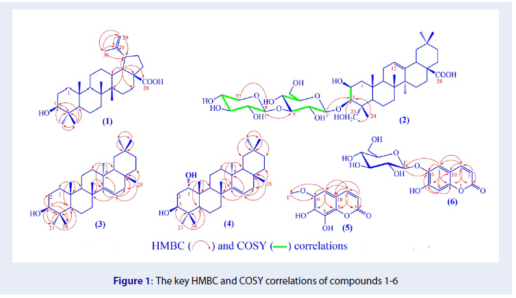
Results: Four triterpenoid compounds, including betulinic acid (1), conyzasaponin G (2), taraxerol (3), and taraxer-14-ene- 1a,3b -diol (4), and two coumarins fraxetin (5), and aesculin (6) were identified.

Conclusion: To the best of our knowledge, they have not been reported in the leaves of Sterculia foetida before, and compound 2 was known to present in Sterculia genus for the first time.

INTRODUCTION

Linn. belonging to Sterculiaceae is grown in tropical areas1. Leaves of Linn. are used in traditional medicine as an aperient, diuretic, and insect repellent2, 3. There were some previous studies on chemical constituents of different parts of this species, which reported the presence of steroids, flavonoids, phenolic, coumarins, phenylpropanoids, and cerebrosides2, 3, 4, 5. Previously, we reported the isolation of the organic compounds from leaves of this species collected in Binh Thuan province, including some triterpenoids, quercetin derivatives, and phenolic compound6, 7. Herein, the continuous chemical study on the hexane and ethyl acetate extracts of leaves was discussed.

MATERIALS AND METHODS

General experimental procedures

The NMR spectra were obtained on a BRUKER AC 500 spectrometer (500 MHz for H-NMR and 125 MHz for C-NMR). The SCIEX X500 QTOF and X500 QTOF machines were used to record the high resolution-mass spectra and the ESI/APCI mass spectra, respectively. Column chromatography was applied to the silica gel normal-phase (Kieselgel 60, 230-400 mesh, Merck). The thin-layer chromatography technique was done by using silica gel plates (Kieselgel 60 F, 0.25 mm, Merck).

Plant material

The leaves were collected in Binh Thuan Province in October 2017. The scientific name of this plant was determined by botanist Dr. Dang Van Son, Institute of Tropical Biology. A voucher specimen, coded No.SFC/TUYEN-1017A, was deposited at the laboratory of Faculty of Environmental Science, Saigon University.

Extraction and isolation

The extraction procedure to obtain 1830 g crude extract by maceration method for three times (3 x 40 L ethanol) at room temperature and partitioned extracts including 450 g of hexane, 650 g of chloroform, 30g of ethyl acetate extracts, and the remaining layer by liquid-liquid partition method was shown more detail in 7.

From the hexane extract (450.5 g), seven fractions were separated by silica gel column chromatography and eluted with hexane-ethyl acetate (1:0, 3:1, 1:1, 1:3, 0:1, v/v, respectively). Fraction SFH.III (60.0 g) was subjected to silica gel column chromatography, eluted with hexane-ethyl acetate (20:1, 9:1, 4:1, 3:2, 1:1) to yield four subfractions. Subfraction SFH.III.2 (18.0 g) was rechromatographed on silica gel column eluting with hexane-ethyl acetate (20:1, 10:1, 5:1) and repeated this process three times to obtain 3 (15.0 mg). The same procedure was applied to subfraction SFH.III.4 (15.2 g) to yield 4 (25.3 mg). Fraction SFH.IV (75.0 g) was applied to a silica gel column and eluted with hexane: ethyl acetate (10:1, 5:1, 3:1, 1:1) to afford four subfractions. Subfraction SFH.IV.3 (20.8 g) was applied to silica gel column and eluted with a solvent system of -hexane-ethyl acetate (3:1) and repeated this process three times on subfractions containing the main compound of SFH.IV.3 checked by TLC plates to obtain 1 (4.5 mg).

The ethyl acetate extract (30.1 g) was separated into five fractions, coded EA.I-EA.V, by silica gel column chromatography and eluted with hexane-ethyl acetate (1:1, 1:3, v/v, respectively) and then chloroform-methanol (9:1, 4:1, 1:1, 0:1, v/v, respectively). Fraction EA.IV (5.3 g) was subjected to silica gel column chromatography and eluted with solvent systems of chloroform-methanol (1:0, 9:1, 4:1, 1:1) and further rechromatographed twice with the same procedure to give 2 (5.0 mg), 5 (4.1 mg), and 6 (5.2 mg).

Figure 1

The key HMBC and COSY correlations of compounds 1-6

RESULTS

The chemical investigation on the leaves collected in Binh Thuan Province led to the isolation of six compounds by the use of efficient separation techniques. From the hexane extract, three compounds 1, 3, and 4 were isolated, while 2, 5, and 6 were isolated from ethyl acetate extract. Their C-NMR data were performed in Table 1, and the following data were H-NMR data.

DISCUSSION

Compound 1 was obtained as a white amorphous powder. Its molecule was determined as CHO by the pseudo molecular ion peak at at 455.3512 (calcd. for [CHO–H], 455.3525). Its C-NMR spectrum showed 30 carbon signals of a triterpene skeleton whose most of the carbon signals resonated in the high magnetic zone from 14 to 56 ppm. An oxygenated methine signal at δ 79.0 was assigned to C-3 as usual. Two signals at δ 150.4 (>C=) and 109.7 (CH=) were characteristic for C-20 and C-29 of a double bond in lupane skeleton. The lowest magnetic signal at δ 179.4 belonged to a carboxyl group. These corresponded to the presence of two broad-singlet proton signals, each integrated 1 proton, at δ 4.61 (H-29a), and 4.74 (H-29b) in H-NMR spectrum. These protons displayed HMBC cross-peaks to methine carbon (46.9, >CH-19), quarterany olefin carbon (150.4, C-20), and methyl carbon (19.4, CH-30). Therefore, the presence of isopropenyl group in lupane skeleton was proved. Five rest singlet methyl proton signals from 0.76 to 0.98 ppm were assigned to H-23 to H-27. The -position of the oxygenated methine proton was confirmed by the signal at δ 3.19 (1H, 11.5, 5.0), since the hydroxy group at C-3 was determined as - or β- position. Based on the above analysis, 1 was elucidated as betulinic acid8.

The NMR analysis of 2 and 1 showed that 2 had one more hydroxyl group at C-2, one more hydroxymethylene instead of the methyl group, and two more sugar units. Two oxymethylene protons presented as two doublet signals at δ 3.63 (1H, 10.5, H-23a), 3.27 (1H, 10.5, H-23b) which had the same HSQC cross-peaks with carbon at δ 65.7 and HMBC correlations with carbons C-3, C-4, C-24/C-23 since these two protons were H-23 or H-24. The chemical shift values of two methyl groups at C-23 and C-24 were around 28 and 15 ppm, respectively, as usual. In the case of 2, one methyl was changed into oxymethylene group, and the rest methyl one was observed at 14.7 ppm, which confirmed the position of the hydroxyl group at C-23. The presence of signals at δ 4.34 (1H, , H-2) and δ 71.2 (C-2) of oxymethine group at C-2 which was determined via COSY correlation between H-2 and H-3. The proton H-2 in compound 2 appeared as a broad-singlet signal at δ 4.34 which determined the -position of this proton or β-OH group at C-2. The NMR spectra of 2 showed two anomeric proton signals at δ 4.51 (1H, , 7.5, H-1ʹ), 4.52 (1H, , 7.5. H-1ʹʹ) and a serial carbinol signals from 3.2 to 3.9 ppm as well as two anomeric carbons at δ 105.2 and 106.0 and nine oxycarbon signals from 62 to 88 ppm which determined the presence of one hexose and one pentose. The COSY spectrum revealed the connection of H-1ʹ/H-2ʹ/H-3ʹ, H-4ʹ/H-5ʹ/H-6ʹ, H-1ʹʹ/H-2ʹʹ/H-3ʹʹ/H-4ʹʹ/H-5ʹʹ. The large coupling constant around 7.5 to 9.0 Hz of H-1ʹ, H-2ʹ, H-4ʹ, H-1ʹʹ, H-4ʹʹ assigned the position of all methine protons of β-glucopyranosyl and β-xylopyranosyl units. The positions of two glucopyranosyl and xylopyranosyl were determined at C-3 and C-3ʹ, respectively, by HMBC correlations of proton H-1ʹ with carbon C-3, of proton H-1ʹʹ with C-3ʹ. The HR-IDA-MS of 2 showed pseudo molecular ion peak at 781.4385 (calcd. for [CHO-H], 781.4374). Based on the above analysis as well as the comparison NMR data of 2 with those reported in the literature9, 2 was identified as conyzasaponin G.

The APCI-MS of 3 and 4 revealed the molecular ion peak at 409.36 [M–HO+H] and 477.47 [M +2HO–H], respectively. NMR data of 3 and 4 displayed signals of triterpenes. The C-NMR showed a pair of signals at around 158 and 117 ppm of a double bond at C-14/C-15 in taraxerane skeleton, which corresponded to the presence of eight singlet proton signals of eight quaternary methyl groups in the range from 0.80 to 1.10 ppm. The C-NMR of 3 showed one oxymethine carbon at δ 79.0 of C-3 as usual, while that of 4 revealed two oxymethine signals at δ 72.9 (C-3) and 71.8 (C-1). The HMBC spectra of 3 and 4 showed cross-peaks of proton H-23 and H-24 with carbon oxymethine C-3. Moreover, HMBC of 4 displayed a correlation of H-25 with the rest oxymethine at 71.8 ppm. Therefore, the positions of two hydroxyl groups in 4 were determined. The proton H-3 appearing as a signal with a large coupling constant of about 11.0 Hz assigned the 3-β-OH whereas the small coupling constant with 3.0 Hz of H-1 in compound 4 suggested the 1-α-OH. The comparison NMR data of 3 and 4 with those reported in the literature, their structures were assigned as taraxerol10 and taraxer-14-ene-1α,3β-diol11.

NMR spectra of compound 5 showed signals of a coumarin skeleton, including 9 carbon signals from 101 to 161 ppm, in which 3 methine carbons showed HSQC cross-peaks with three proton signals at δ 6.16 (1H, , 9.5, H-3), 7.81 (1H, , 9.5, H-4), and 6.77 (1H,, H-5). The HMBC correlations of these protons (as shown in Figure 1) were used to determine their positions. At a higher magnetic field, the NMR spectra of 5 showed signals at δ 3.87 (3H, , OCH) and δ 56.7 (OCH). This methoxy group was attached to C-6, which was determined via HMBC cross-peaks between methoxy proton and carbon C-6. The ESI-MS of 5 showed pseudo molecular ion peak at 209.06 [CHO+H]. Compound 5 was identified as fraxetin via the good compatible NMR data of 5 with the reported data12.

The comparison NMR spectra of 5 and 6 showed that 6 was a coumarin glycoside. This was proved by the presence one more sugar unit and one less methoxy group. The proton NMR of 6 showed a set of signals including δ [4.84 (1H, , 7.5, H-1ʹ) and 3.4-3.9 ()] and δ [104.7 (C-1ʹ) and 62.5-78.2]. The constant coupling analysis of proton H-1ʹ (, 7.5) and H-4ʹ ( 9.0) suggested that all methine protons of the sugar unit were positions. The HMBC correlation of the anomeric proton H-1ʹ and carbon C-6 as well as all other HMBC correlations (as shown in Figure 1) confirmed the structure of 6. The HR-IDA-MS of 6 showed a signal at 339.0720 (calcd. for [CHO-H], 339.0716). Based on the above analysis, the structure of 6 was determined as aesculin13.

CONCLUSION

In the continuous study on the leaves of Linn. collected in Binh Thuan province, four triterpenoids and two coumarin derivatives were isolated, including betulinic acid (1), taraxerol (3), and taraxer-14-ene-1,3-diol (4) isolated from the hexane extract and conyzasaponin G (2), fraxetin (5), and aesculin (6) isolated from the ethyl acetate extract. Their chemical structures were determined by the NMR, MS data analysis, and their spectroscopic data and physical properties with those reported in the literature.

ABBREVIATIONS

HR-IDA-MS: High resolution-Information dependent acquisition-Mass spectrometry

ESI/APCI-MS: Electrospray ionization/Atmospheric pressure chemical ionization-Mass spectrometry

H NMR: Proton nuclear magnetic resonance

C NMR: Carbon-13 nuclear magnetic resonance

COSY: Correlation spectroscopy

HSQC: Heteronuclear single quantum coherence

HMBC: Heteronuclear multiple bond correlation

s: singlet

: broad singlet

: doublet

: doublet of doublets

: multiplet

- triplet-like

COMPETING INTEREST

The authors declare no competing financial interest.

AUTHORS’ CONTRIBUTION

Pham N.K.T has contributed in conducting experiments, acquisition of data, and interpretation of data. Nguyen T. Q. T., Huynh C. D., Pham D. T., Nguyen T. D., Tran D. D. C., Huynh B. L. C., Nguyen T. A. T. interpreted NMR and MS data as well as searched the bibliography. Nguyen K. P. P. and Nguyen T. H. T gave final approval of the manuscript to be submitted.

ACKNOWLEDGMENT

Sai Gon University funded for this research under grant number CS2020-50.

Table 1

13C-NMR data of six isolated compounds

No

1c

2m

3c

4a

5a

6a

1

38.8

44.4

37.7

71.8

2

27.4

71.2

27.1

34.7

161.0

161.1

3

79.0

84.0

79.0

72.9

113.3

113.6

4

38.9

43.1

38.7

39.6

145.3

144.6

5

55.4

49.5

55.5

48.7

101.2

117.8

6

18.3

18.6

18.8

19.3

146.2

143.5

7

34.4

33.9

41.3

41.9

140.2

153.0

8

40.8

40.6

38.9

39.6

133.6

104.3

9

50.6

48.2

49.3

41.1

139.7

152.7

10

37.3

37.5

37.5

42.3

111.7

112.3

11

20.9

24.7

17.5

17.2

12

25.6

123.6

37.7

33.8

13

38.4

145.4

35.7

38.4

14

42.5

43.2

158.1

159.6

15

30.6

28.8

116.8

117.2

16

32.2

24.1

36.6

38.3

17

56.3

47.3

38.0

36.5

18

49.3

42.8

48.7

49.6

19

46.9

47.3

35.1

37.3

20

150.4

31.6

28.8

29.9

21

29.7

35.0

33.7

33.8

22

37.0

33.5

33.1

35.4

23

28.0

65.7

28.0

28.5

24

15.4

14.7

15.4

15.9

25

16.1

17.5

15.4

16.8

26

16.1

17.9

29.8

26.4

27

14.7

26.5

25.9

21.6

28

179.4

174.6

29.9

30.1

29

109.7

33.6

33.3

33.5

30

19.4

24.0

21.3

30.2

105.2

56.7

104.7

74.7

74.6

88.0

78.2

69.4

71.3

77.4

77.5

62.2

62.5

1ʹʹ

106.0

2ʹʹ

75.3

3ʹʹ

77.7

4ʹʹ

71.0

5ʹʹ

67.1

References

  1. . Vo VC. Dictionary of medical plants in Vietnam. Medical Publishing House. 2002; : 1083.
  2. . Xia PF, Feng ZM, Yang YN, Zhang PC. Two flavonoid glycosides and a phenylpropanoid glucose ester from the leaves of Sterculia foetida. Journal of Asian Natural Products Research 2009; 11(8): 766-771.
  3. . Mujumdar AM, Naik DG, Waghole RJ, Kulkarni DK, Kumbhojkar MS. Pharmacological studies on Sterculia foetida leaves. Pharmaceutical Biology 2000; 38(1): 13-17.
  4. . Anjaneyulu ASR, Murty VS. Two rare tetramethyl ethers of quercetin from Sterculia foetida Linn. Indian Journal of Chemiscal Section B 1981; 20(1): 87-88.
  5. . Kale SS, Darade V, Thakur HA. Analysis of fixed oil from Sterculia foetida Linn. International Journal of Pharmaceutical Sciences and Research 2011; 2(11): 2908-2914.
  6. . Pham DT, Doan TDC, Nguyen TP, Mai DT, Pham NKT, Nguyen KPP. Quercetin derivatives from the ethyl acetate of the leaves of Sterculia foetida Linn. Vietnam Journal of Chemistry 2018; 56(4e): 120-123.
  7. . Pham NKT, Nguyen TD, Doan TDC, Ha TD, Tran NMA, Tran TD, Mai DT, Nguyen TP. Stercufoetin A, new oleanane-type triterpenoid from the leaves of Sterculia foetida L. Natural Product Research. 2020; :
  8. . Satiraphan M, Pamornsilpadaharm P, Sittisombut C, Sotanaphun U, Raynaud F, Garbay C, Michel S, Cachet X. Lupane triterpenes from the leaves of the tropical rain forest tree Hopea odorata Roxb. and their cytotoxic activities. Biochemical Systematics and Ecology 2012; 44: 407-412.
  9. . Su Y, Guo D, Guo H, Liu J, Zheng J, Koike K, Nikaido T. Four new triterpenoids from Conyza blinii. Journal of Natural Products 2001; 64(1): 32-36.
  10. . Xu R, Fazio GC, Matsuda SPT. On the origins of triterpenoid skeletal diversity. Phytochemistry 2004; 65: 261-291.
  11. . Li S, Shi Y, Shang XY, Cui BS, Yuan Y, Chen XG. Triterpenoids from the roots of Pterospermum heterophyllum Hance. Journal of Asian Natural Products Research 2009; 11(7): 652-657.
  12. . Yasuda T, Fukui M, Nakazawa T, Hoshikawa A, Ohsawa K. Metabolic fate of fraxin administered orally to rats. Journal of Natural Products 2006; 69(5): 755-757.
  13. . Yu M, Sun A, Zhang Y, Liu R. Purification of coumarin compounds from Cortex fraxinus by adsorption chromatography. Journal of Chromatographic Science 2014; 52: 1033-1037.

Comments