

Synthesis and optical properties of ZnS nanoparticles decorated on SiO2 nanospheres
- Faculty of Physics, VNU University of Science, Vietnam National University - Hanoi
Abstract
Introduction: In this study, ZnS nanoparticles were decorated on SiO2 nanospheres via coprecipitation and the Stӧber method.
Methods: The materials were studied by X-ray diffraction (XRD), nano scanning electron microscopy (SEM), and photoluminescence spectra (PL).
Results: ZnS nanoparticles exhibit a sphalerite structure with an average particle size of 2.8 nm and SiO2 in the form of 115 nm nanospheres. The absorption spectrum of these nanoparticles displays a prominent band peaking at 320 nm. Additionally, at 300 K, the PL spectrum of ZnS reveals a broad band, comprising two component bands with peaks at 470 nm and 491 nm, attributed to lattice defects such as zinc and sulfur vacancies, interstitials, and surface states. Upon decorating ZnS nanoparticles on the surface of SiO2 nanospheres, the UV‒Vis absorption and PL spectra of ZnS shift toward longer wavelengths. The maximum peak of the UV‒vis spectrum shifted from 322 to 328 nm, and two bands in the PL spectrum slightly changed to 472 and 494 nm.
Conclusion: The shifts in the UV‒vis and PL spectra are due to alterations in the surface states of the ZnS nanoparticles induced by the presence of SiO2. From the dependence of the PL spectra of ZnS nanopartcles and SiO2-decorated ZnS on the measured temperature from 10 to 300 K, the activation energies of ZnS and SiO2-decorated SiO2 were estimated to be 38 and 40 meV, respectively.
INTRODUCTION
Semiconductor nanomaterials have garnered extensive attention due to their vast potential applications, including catalysis, photonics, nonlinear optical devices, light-emitting diodes, flat displays, and infrared windows 1, 2, 3, 4, 5, 6. Nanostructured materials exhibit novel optical properties owing to the quantum size effect, making the precise control of their size a significant challenge in research and manufacturing. ZnS is one of the most critical and representative semiconductors, prompting various efforts to manipulate its optical properties. Techniques such as doping with elements such as Mn and Cu, employing polymer capping, and fabricating core/shell structures with materials such as ZnO and SiO have been explored7, 8, 9, 10, 11, 12, 13. Silicon dioxide (SiO)-based core shell particles have been widely studied because of their chemical inertness, ability to act as stabilizers, and ability to prevent particle coalescence. Their nanostructures can be homogeneously prepared and uniformly dispersed in different solutions. Due to its high biocompatibility and functionalized surface, SiO readily binds to pigments, metal ions, and biomolecules14. Combining ZnS with SiO holds tremendous potential for applications in photocatalysis, environmental treatment, and enhancing PL 15.
SiO@ZnS core-shell nanoparticles have been synthesized by various reported methods. For example, SiO@ZnS core-shell nanoparticles were synthesized by a thermal decomposition approach by Jatin Mahajan et al.15. Ethiraj et al. synthesized SiO@ZnS with thioglycerol molecules attached to functionalised silica particles16. Dhas et al. synthesized SiO@ZnS core-shell nanoparticles using ultrasonic irradiation 17, and Velikov et al. synthesized fluorescein isothiocyanate-incorporated SiO@ZnS core-shell nanoparticles by combining homogeneous precipitation and thermal decomposition methods18. These reports analyzed the microstructure, morphology and UV‒Vis absorption spectra of SiO@ZnS core-shell nanoparticles, but studies on the luminescence spectra have not been discussed in detail.
With the process of ZnS decoration on SiO, defects and surface states in ZnS will be generated; therefore, the crystal structure and optical properties of ZnS will be affected. In this paper, we present the facile synthesis of ZnS nanoparticles decorated on SiO nanospheres and demonstrate the influence of SiO on the crystal structure, UV‒Vis spectrum and PL spectra of ZnS nanoparticles from 10 to 300 K.
MATERIALS AND METHODS
Tetraethoxysilane (TEOS), Si(OC2H5)4 (99,98%), (3-aminopropyl) trimethoxysilane (APTMS) (99,00%), zinc acetate Zn(CHCOO),2 H2O (99,99%), thioacetamide (TAA), CH3CSNH2 (99%), ammonium NH3 (25%), and absolute ethanol were purchased from Sigma Aldrich, China. All chemicals used were of analytical grade. Deionized (DI) water was used in all experiments.
ZnS nanoparticles were synthesized by a coprecipitation method from solutions of 0.01 M TAA and 0.01 M zinc acetate according to the following process: 30 ml of 0.01 M TAA solution was slowly added to 30 ml of 0.01 M zinc acetate solution, and the mixture was stirred for 2 hours to obtain a white precipitate. The white precipitate was centrifuged and filtered 3 times and then dried at 80°C for 15 hours to obtain ZnS powder. SiO2 nanospheres were synthesized by the Stӧber method from TEOS, NH3 and absolute ethanol. ZnS (0.02 g), SiO2 (0.1 g), APTMS (0.2 ml), DI water (40 ml) and absolute ethanol (10 ml) were mixed with magnetic stirring for 5 hours. The mixture was centrifuged and dried at 80°C for 15 hours to obtain ZnS nanoparticles decorated on SiO2 nanospheres (denoted as SiO2@ZnS).
RESULTS AND DISCUSSION
Structure and morphology of ZnS nanoparticles and SiO@ZnS
Figure 1a shows the XRD pattern of the ZnS nanoparticles (NPs). The diffraction peaks at 2 23.91, 48.11, and 57. are attributed to the (111), (220), and (311) atomic planes of ZnS, respectively (according to PDF No. 96-500-0089). The existence of these peaks indicates that as-ZnS is a single–phase, cubic structure belonging to the symmetry group. The XRD pattern of SiO@ZnS also shows peaks at 2 23.91, 48.11, and 57.11°, similar to the XRD pattern of ZnS nanoparticles (Figure 1b). This indicates that as-SiO does not affect the structure of ZnS when ZnS is decorated on SiO. From the XRD patterns and the Debye–Scherrer formula, where D is the crystalline size, λ is the wavelength of CuKα radiation (1.54056 Ǻ), β is the full width at half maximum and θ is the Bragg diffraction angle, the crystalline size of ZnS was calculated to be approximately 2.8 nm using a highly intense Bragg peak at 2 23..

XRD pattern of ZnS nanoparticles (a) and ZnS nanoparticles decorated on SiO2 nanospheres (b)
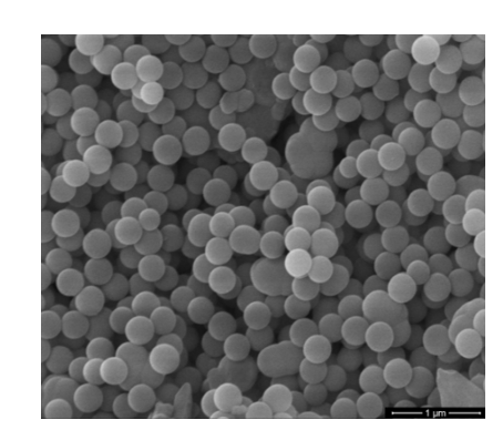
Figure 2 displays an SEM image of SiO. The image reveals that SiO exhibits a spherical morphology and uniform distribution with an average particle size of 115 nm (as depicted in Figure 3).

SEM image of SiO2 nanospheres

Particle size distribution histogram of SiO2 nanospheres
SEM images of the ZnS nanoparticles showed that the ZnS nanoparticles aggregated with each other in the clusters (Figure 4). However, in the SEM image of SiO@ZnS, the ZnS nanoparticles agglomerated on the surface of the SiO nanospheres (Figure 5). Hence, SiO nanospheres acted as templates for ZnS nanoparticle anchoring.

SEM image of ZnS nanoparticles

SEM image of S ZnS nanoparticles decorated on SiO2 nanospheres
UV‒Vis absorption spectra of ZnS nanoparticles and SiO@ZnS
Figure 6 shows the UV‒Vis absorption spectra of the ZnS nanoparticles (a) and SiO@ZnS (b). In the UV‒Vis absorption spectrum of ZnS (Figure 6a), a prominent band appears with a maximum peak at 322 nm (3.85 eV), attributed to the near-band-edge absorption of ZnS 19, 20. This peak's maximum value demonstrates a blueshift compared to the 340 nm (3.65 eV) absorption peak of cubic bulk ZnS. Upon the addition of ZnS to the SiO nanospheres, the absorption peak shifts to a longer wavelength of 328 nm (Figure 6b).

UV- Vis absorption spectra of ZnS nanoparticles (a) and ZnS nanoparticles decorated on SiO2 nanospheres (b)
PL spectra of ZnS nanoparticles and SiO@ZnS
Figure 7 presents the PL spectra of both ZnS nanoparticles and SiO@ZnS when excited by 325 nm radiation from a He-Cd laser at room temperature.

PL spectra of ZnS nanoparticles (a) and ZnS nanoparticles decorated on SiO2 nanospheres (b) at 300 K
At 300 K, there appears to be a broad luminescent band including two component bands at 470 and 491 nm (Figure 7a). These bands are attributed to defects in the crystal lattice, such as vacancies of zinc, sulfur, interstitial atoms of zinc, sulfur, and surface states 21, 22. When ZnS was decorated on SiO, the intensity of these bands decreased significantly, while the peak positions of the two component bands shifted toward longer wavelengths at 472 and 494 nm (Figure 7b).
From the above results, we suppose that as-SiO attaches to ZnS by the -NH amin of APTMS; in addition to ZnS decorating SiO, ZnS nanoparticles agglomerate together. Hence, the particle size of ZnS increases, leading to a shift in the UV‒Vis absorption and PL spectra of SiO@ZnS toward longer wavelengths.
At 10 K, in the PL spectrum of the ZnS nanoparticles, there is also a broad band with two component bands at 464 and 490 nm assigned to defects in the ZnS crystal lattice, similar to that at 300 K. For SiO@ZnS, at 10 K, there are bands at 440 and 482 nm, in which the 440 nm band has a strong intensity and the 482 nm band appears weakly on the right side of the 440 nm band. When the measurement temperature was increased from 10 K to 300 K, the position of the PL bands shifted toward longer wavelengths, and their intensity decreased rapidly (approximately 50 times) with temperature (Figure 8, Figure 9). The redshift of the PL spectra due to the energy band gap decreases with increasing temperature owing to exciton–phonon coupling and lattice deformation 23, 24.
The dependence of the luminescence intensity on temperature is as follows:
in which I is the PL intensity at 10 K
A is a constant
E is the activation energy
k is the Boltzmann constant

Temperature-dependent PL spectra of ZnS nanoparticles when changing the measurerement temperature from 300 to 10K

Temperature-dependent PL spectra of ZnS nanoparticles decorated on SiO2 nanospheres when changing the measurement temperature from 300 to 10 K
From the dependence of the PL intensity on the measurement temperature (10/T), the activation energies for ZnS (at 464 nm) and SiO@ZnS (at 440 nm) were calculated to be 38 and 40 meV, respectively (refer to the inset in Figure 8 and Figure 9). These values are in agreement with references 25, 26. From the above results, it can be inferred that upon decorating ZnS on SiO spheres, the surface states of ZnS undergo changes, resulting in subtle variations in both the UV‒Vis and PL spectra, as well as the activation energy of ZnS.
CONCLUSION
ZnS nanoparticles and ZnS nanoparticles decorated on SiO nanospheres were successfully fabricated. The cubic-structured ZnS nanoparticles, averaging 2.8 nm in crystalline size, were decorated on SiO nanospheres. Upon attachment of ZnS onto SiO spheres, both the UV‒Vis absorption and luminescence spectra exhibited a shift toward longer wavelengths, attributed to alterations in the surface states of ZnS. Notably, the luminescence spectra of ZnS and SiO@ZnS increased by 50 times, while the position of the luminescent band shifted slightly when the measurement temperature decreased from 300 K to 10 K. The activation energies for ZnS and ZnS nanoparticles decorated on SiO nanospheres were determined to be 38 and 40 meV, respectively. These results are the basis for our further research on the photocatalysis and luminescence enhancement of ZnS nanoparticles decorated on SiO templates.
ACKNOWLEDGEMENTS
We extend our gratitude to the QG 22.15 project of Vietnam National University, Hanoi, for providing financial support for this paper.
AUTHOR CONTRIBUTIONS
All the authors contributed to the study conception and design. Material preparation, data collection and analysis were performed by Bui Hong Van, Nguyen Thi Bao Yen, Doan Thi Kim Dung, and Hoang Chi Hieu.
The first draft of the manuscript was written by Bui Hong Van, and all the authors commented on previous versions of the manuscript. All the authors have read and approved the final manuscript.
COMPETING INTERESTS
The authors declare that they have no known competing financial interests or personal relationships that could have appeared to influence the work reported in this paper.