

Molecular cloning and isolation of a recombinant alpha-Momorcharin in E. coli against Pyricularia oryzae
- Ho Chi Minh City Open University, Vietnam
Abstract
Introduction: Alpha-Momorcharin (α-MMC) is a member of the ribosome-inactivating protein (RIP) family that has been widely used as an antitumor, antiviral and antifungal agent.
Methods: In this study, the codons of DNA encoding α-MMC were optimized for expression in E. coli and cloned into the pET-28a(+) vector. The protein was then expressed in E. coli strain BL21 (DE3) and purified by nickel affinity chromatography.
Results: Under IPTG induction, α-MMC was expressed at approximately 50% of the total protein, showing high-level recombinant protein expression in E. coli. A high amount of purified α-MMC (70 mg) was isolated from 1 L LB culture medium of E. coli BL21 (DE3) with approximately 95% purity. Interestingly, α-MMC inhibited the mycelial growth of Pyricularia oryzae in a concentration-dependent manner.
Conclusion: Using a microbial system for α-MMC expression provides a promising method for the design of a new agent against pathogens.
INTRODUCTION
Ribosome-inactivating proteins (RIPs) are found in many plant species within different tissues. RIPs can act as protein synthesis inhibitors because of their toxicity with the ability to biologically inhibit the synthesis of proteins based on N-glycosidase activity 1. The potential applications of RIPs have been widely studied in both medicine and agriculture (Figure 1)2, 3. In medicine, RIPs are fused with antibodies or other carriers to generate immunotoxins or toxic agents to cancer cells2, 4, 5, 6. In agriculture, RIPs are expressed in plants under stressful conditions to resist infection by pathogens such as viruses, fungi and insects7, 8, 9, 10, 11.
Alpha-Momorcharin (α-MMC) belongs to a member of the ribosome-inactivating protein (RIP) family, which is abundantly found in the seeds of Momordica charantia 12, 13, 14.The α-MMC protein has various important agricultural activities, such as antifungal and antiviral activities 7, 15, 16. Insight into the mechanism of α-MMC against fungal and viral pathogens has been studied. α-MMC can directly inhibit pathogens by entering the cell wall through the endoplasmic reticulum, leading to inhibition of protein synthesis, thereby inhibiting fungal growth and killing fungal cells16. However, there is increasing evidence that the main cause of toxicity in germ cells is their ability to induce reverse inhibition of apoptosis. α-MMC inhibits the synthesis of protein by hydrolytically removing a specific residue of adenine from a single-stranded loop of rRNA 1. The hydrolysis of the N-glycosidic bond takes place on the highly conserved Sarcin/Ricin Loop region of the rRNA, which interrupts the interaction of elongation factor II (EF-2), causing the ribosome to fail to bind and ultimately inhibit protein synthesis at the translation step, causing cell death 17.

Ribosome-inactivating proteins (RIPs) have antitumor, anti-inflammatory, antifungal and antiviral activities.
Many studies have demonstrated that the α-MMC protein plays an important role in many fields. The most promising application of α-MMC in medicine is in the prevention of cancer. α-MMC has also been shown to inhibit the growth of breast cancer cells through inhibition of tumor growth and induction of apoptosis6, 18, 19, 20, 21, 22. In addition, α-MMC is a potential therapeutic agent for HIV/AIDS via its ability to inhibit HIV replication in acutely and chronically infected cells23, 24. In agriculture, the α-MMC protein showed a wide range of antiviralactivity (resistant to Chiliveinal mottle virus, Cucumber mosaic virus, Tobacco mosaic virus)16 and antifungal activity (resistant to Fusarium spp., Aspergillus spp., Sclerotinia sclerotiorum, Bipolaris maydis, Magnaporthe grisea) 11. In fact, extraction of α-MMC protein from the seeds of bitter melon by traditional techniques has limited the mass of protein for studying the structure and biological functions of traditional approaches25. In this study, we aimed to clone, express, and purify α-MMC protein with high purity in an E. coli system. The isolated protein was used to resist Pyricularia oryzae. This finding shows great promise for the expansion of potential α-MMC protein applications against pathogens.
MATERIALS AND METHODS
Construction of plasmid
A target α-MMC protein was generated in plasmid pET28a(+). The DNA sequence encoding α-MMC was obtained from GenBank (CAA40869.1). DNA of α-MMC,which was optimized by the codon optimization tool from Integrated DNA Technologies (IDT) for expression in the E. coli system 26, was then synthesized by the Macrogen company (Korea).DNA encoding α-MMC was amplified by PCR using the α-MMC gene (optimal codons, IDT, USA) as the template and the primer pair ONF/ONR (ONF: 5'-cag cca tat gga tgt tag ctt tcg ttt gtc ggg tgc tga t -3'; ONR: 5'- ggt gct cga gtc agt agc tcg aaa agc cat gtg -3'). The pcr product was inserted into treated pET28a(+) (Merck Millipore, Germany) at the NdeI and XhoI sites by T4 DNA ligase, resulting in the generation of the plasmid pET-MMC.
Amino acid sequence of α-MMC protein:
MGSSHHHHHHHHSSGLVPRGSHMDVSFRL
SGADPRSYGMFIKDLRNALPFREKVYNIPLLL
PSVSGAGRYLLMHLFNYDGKTITVAVDVTNV
YIMGYLADTTSYFFNEPAAELASQYVFRDARR
KITLPYSGNYERLQIAAGKPREKIPIGLPALDSA
ISTLLHYDSTAAAGALLVLIQTTAEAARFKYIEQ
QIQERAYRDEVPSLATISLENSWSGLSKQIQLA
QGNNGIFRTPIVLVDNKGNRVQITNVTSKVVT
SNIQLLLNTRNIAEGDNGDVSTTHGFSSY
Expression and purification of proteins
The plasmid pET-MMC encoding the α-MMC protein was chemically transformed into E. coli strain BL21 (DE3). The bacteria were cultured in Luria-Bertani medium (LB) containing 40 mg/L kanamycin, and the E. coli cells were grown at 37 °C with shaking at 200 rpm to reach an OD600 of 0.8-1. IPTG (Sigma Aldrich, St. Louis, USA) was subsequently added to a final concentration of 0.75 mM. The E. coli cells were continuously incubated for 10 hours at 15 °C with shaking at 200 rpm before being harvested by centrifugation. The pellet was then resuspended in lysis buffer consisting of 20 μg/ml DNase I and 1 mM PMS. The cell wall was broken by sonication, and the insoluble cell debris was then removed by centrifugation at 20000 rpm for 1 hour at 4 °C. The soluble fraction was added to a His-tag column (Thermo Fisher Scientific, Waltham, MA, USA). Subsequently, the column was washed with 30 volumes of column with 20 mM Tris-HCl, 100 mM NaCl and 10 mM imidazole buffer, pH 7. The proteins were finally eluted with buffer containing 20 mM Tris-HCl, 100 mM NaCl and various imidazole concentrations. The imidazole chemical in the protein solution was removed by centrifugation using an Amicon Ultra15 centrifugal filter (EMD Milipore). The pure α-MMC protein was collected and analyzed by SDS‒PAGE. Total protein expression was analyzed by AlphaEase software. The pure protein was measured by a NanoDrop spectrophotometer (Thermo Fisher Scientific, USA).
Testing the antifungal activity of α-MMC against pathogens
Initial experiments investigated the antifungal activity against Pyricularia oryzae. The α-MMC protein with different concentration ranges and control (water) samples were evenly added to the surface of the PDA-agar plate. Subsequently, the fungal suspension and control water samples were added into the center of the PDA-agar plate and incubated for 24 hours at 28 °C 27.
RESULTS
Construction of plasmid pET-MMC
The DNA sequence of α-MMC was first optimized for expression in an E. coli system by the codon optimization tool from Integrated DNA Technologies (IDT). The α-MMC gene was provided with the best sequence option by screening and filtering sequences to lower complexity and minimize secondary structures. The α-MMC gene fragment was amplified by PCR with a pair of specific primers ONF and ONR with additional restriction enzyme cutting sites NdeI (forward primer) and XhoI (reverse primer). PCR products were analyzed by gel electrophoresis with 2% agarose, which showed a single bright band near the 800 bp band of the DNA ladder (Figure 2A), corresponding to the theoretical size of the α-MMC gene fragment of 812 bp.
The pET28 plasmid and the α-MMC target gene after treatment with 2 restriction enzymes, XhoI and NdeI, were cloned by T4 DNA ligase. The ligated product was transformed into E. coli strain DH5α and spread on LB-agar medium containing the antibiotic kanamycin. The colonies grown on the medium were selected to clarify the presence of recombinant vector in E. coli strain DH5α by colony PCR. The electrophoresis results showed that 2 colonies (in 7 selected colonies) revealed a band at approximately 1000 bp of the DNA ladder, corresponding to the theoretical size design (1055 bp) (Figure 2B). The recombinant DNA was then sequenced by Macrogen. Inc. (Korea). The sequencing result revealed 100% similarity with the theoretical sequence (data not shown) that clarifies the recombinant vector pET-MMC bearing the α-MMC gene.

DNA electrophoresis.A) PCR product of the
Protein expression and purification
To investigate the expression of the α-MMC protein in a bacterial system, the recombinant plasmid pET-MMC was chemically transformed into the E. coli strain BL21(DE3). The IPTG for induction was added after the cells were grown to reach OD0.8-1. The level of protein expression was analyzed by SDS‒PAGE. The results showed that there was a bright band of protein near the 30 kDa band of the ladder, corresponding to the theoretical molecular weight of the α-MMC protein (31.388 Da) under the addition of IPTG. In contrast, the α-MMC protein was not expressed without the addition of IPTG. Using the AlphaEase FC Software (Alpha Innotech) for calculation of protein expression 28, the α-MMC protein was expressed with a high protein level in E. coli of approximately 50% of total protein under induction of IPTG (Figure 3A).
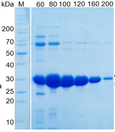
The α-MMC protein was designed with 6xHis for purification. α-MMC fused with a His-tag at the N-terminus was purified by chromatography using the His-tag column. SDS‒PAGE showed that pure α-MMC was collected after elution with different concentrations of imidazole (Figure 3B). The molecular weight of α-MMC is 31.388 Da, which shifted near the 30 kDa band of the ladder. Using the NanoDrop spectrophotometer to measure the protein concentration (total protein fragments with imidazole concentrations of 100 to 200 mM), approximately 70 mg of pure α-MMC was obtained in 1L LB culture medium, which implies that α-MMC can be highly expressed in bacterial systems.

SDS‒PAGE of α-MMC protein. A) Protein expression in
Effect of α-MMC protein on mycelial growth of Aspergillus nomius and Pyricularia oryzae
Theα-MMC protein has previously been shown to confer resistance to fungi by blocking the spread of pathogens. In this study, we tested the antifungal activity of the α-MMC protein against Pyricularia oryzae (causing blast disease). Compared with the control, the difference between treatments with variant α-MMC protein concentrations was evident in the assays. Treatments with 5 µM, 20 µM, 100 µM and 200 µM α-MMC against Pyricularia oryzae (Figure 4) showed that the mycelial growth of fungi was inhibited in a concentration-dependent fashion after 24 hours of incubation. Specifically, for Pyricularia oryzae treated with water as the control, the fungal colonies were brown in color and spread over the plate. At low concentrations of α-MMC (5 µM and 20 µM), the inhibition of fungal growth was still weak, with a diameter of fungal colonies of 3.4 cm. Increasing concentrations of α-MMC at 100 µM and 200 µM clearly showed antimicrobial activity against the blast fungus with a colony diameter of 1.75 cm and nonfungal growth, respectively (Figure 4).

Testing of antifungal activity of protein α-MMC. Growth inhibition of
DISCUSSION
RIPs (ribosome-inactivating proteins) have attracted the attention of biologists in the field of biomedical research because of their diverse activities, including anticancer, antiviral and anti-inflammatory effects. Many studies have demonstrated that RIPs are potent protein toxins with diverse biopharmacological properties, notably rRNA N-glycosidase activity1. α-MMC is a member of the RIP family that has been extracted from the seeds of Momordica charantia and plays a role in antitumor, antiviral, and antifungal activities. However, the biological functions of α-MMC are still not fully understood, partly because of the difficulty in purifying this protein from seeds with traditional approaches25. Mass production of α-MMC is needed for further studies of functional analysis and applications. Using the E. coli system for the expression of α-MMC has been considered an efficient approach for producing high amounts of protein. Herein, optimization of codons for expression in the E. coli system allows lower complexity and minimizes secondary structures. The expression of α-MMC in E. coli strain BL21 (DE3) was approximately 50% of the total proteins under the induction of IPTG, indicating high-level recombinant protein expression in the bacterial system. Using the His-tag column, approximately 70 mg of α-MMC with 95% purity was isolated from 1 L LB culture medium. This shows that α-MMC is expressed in the soluble phase and can easily be purified for functional analysis. Preliminary data showed that α-MMC was able to inhibit the mycelial growth of Pyricularia oryzae in a concentration-dependent fashion. This is in line with a previous study of α-MMC activity against other fungi, such as Fusarium solani and Fusarium oxysporum11. The fungal-inhibiting activity of α-MMC may be involved in RNA N-glycosidase and nuclease activities 11.
CONCLUSIONS
In this study, the pET-MMC vector carrying an α-MMC target gene was genetically generated. This recombinant vector was able to be expressed in an E. coli system with a high amount of α-MMC protein in the presence of the inducer IPTG. The α-MMC protein was homogeneously isolated by chromatography using a His-tag column. Preliminary results showed that α-MMC had activity against Pyricularia oryzae. This is the basis for further studies, including detailed evaluation of the growth inhibition of mycelium for investigation of the anticancer, anti-inflammatory, antifungal and antiviral activities.
COMPETING INTERESTS
There is no conflict of interest.
AUTHORS' CONTRIBUTIONS
D.T.D designed and T.T.T.N performed all experiments. T.T. and D.T. wrote the paper.
ACKNOWLEDGMENTS
We would like to thank Dr. Le Thi Truc Linh and Ms. Duong Nhat Linh (Ho Chi Minh City Open University, Vietnam) for kindly providing fungi.
ABBREVIATIONS
α-MMC: Alpha-Momorcharin
E. coli: Escherichia coli
EF-2: Elongation factor II
IDT: Integrated DNA Technologies
IPTG: Isopropyl β- d-1-thiogalactopyranoside
LB: Luria-Bertani medium
PCR: Polymerase chain reaction
PDA: Potato Dextrose Agar
RIPs: Ribosome-inactivating proteins
SDS‒PAGE: Sodium dodecyl-sulfate polyacrylamide gel electrophoresis